
Page 120 - The Netter Collection of Medical Illustrations - Integumentary System_ Volume 4 ( PDFDrive )
P. 120
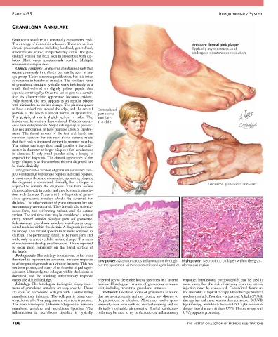
Plate 4-35                                                                                            Integumentary System


       GRANULOMA ANNULARE


       Granuloma annulare is a commonly encountered rash.
       The etiology of this rash is unknown. There are various                                 Annulare dermal pink plaque.
       clinical presentations, including localized, generalized,                               Typically asymptomatic and
       subcutaneous, actinic, and perforating forms. The gen-                                  undergoes spontaneous resolution
       eralized version has been seen in association with dia-
       betes.  Most  cases  spontaneously  resolve.  Multiple
       treatment strategies exist.
         Clinical Findings: Granuloma annulare is a rash that
       occurs  commonly  in  children  but  can  be  seen  in  any
       age group. There is no race predilection, but it is twice
       as common in females as in males. The localized form
       of granuloma annulare typically starts insidiously as a
       small,  flesh-colored  to  slightly  yellow  papule  that
       expands centrifugally. Once the lesion gets to a certain
       size,  its  characteristic  appearance  becomes  evident.
       Fully  formed,  the  area  appears  as  an  annular  plaque
       with minimal to no surface change. The plaque appears
       to have a raised rim around the edge, and the central   Generalized
       portion of the lesion is almost normal in appearance.   granuloma
       The  peripheral  rim  is  slightly  yellow  in  color.  The   annulare
       lesions  can  be  entirely  flesh  colored.  Patients  experi-  in a child
       ence minimal symptoms. Slight itching may be present.
       It is not uncommon to have multiple areas of involve-
       ment.  The  dorsal  aspects  of  the  feet  and  hands  are
       common  locations  for  this  rash.  Some  patients  relate
       that their rash is improved during the summer months.
       The lesions can range from small papules a few milli-
       meters in diameter to larger plaques a few centimeters
       in  diameter.  If  only  small  papules  exist,  a  biopsy  is
       required for diagnosis. The clinical appearance of the
       larger plaques is so characteristic that the diagnosis can
       be made clinically.
         The generalized version of granuloma annulare con-
       sists of numerous widespread papules and small plaques.
       In most cases, there are no annulare-appearing plaques;
       the  diagnosis  is  considered  clinically,  but  a  biopsy  is                                 Localized granuloma annulare
       required  to  confirm  the  diagnosis.  This  form  occurs
       almost exclusively in adults and may be seen in associa-
       tion with diabetes. Patients with a diagnosis of gener-
       alized  granuloma  annulare  should  be  screened  for
       diabetes. The other variants of granuloma annulare are
       uncommonly encountered. They include the subcuta-
       neous  form,  the  perforating  variant,  and  the  actinic
       variant. The actinic variant may be considered a unique
       entity,  termed  annular  elastolytic  giant  cell  granuloma.
       Subcutaneous  granuloma  annulare  manifests  as  deep-
       seated nodules within the dermis. A diagnosis is made
       via biopsy. This variant appears to be more common in
       children. The perforating variant is the rarest form and
       is the only variant to exhibit surface change. The areas
       of involvement develop small erosions. This is reported
       to  occur  most  commonly  on  the  dorsal  surface  of
       the hands.
         Pathogenesis: The etiology is unknown. It has been
       theorized to represent an abnormal immune response   Low power. Granulomatous inflammation through-  High power. Necrobiotic collagen within the gran-
       to a foreign antigen such as a virus or bacteria. This has   out the specimen with necrobiotic collagen bundles  ulomatous region
       not been proven, and many other theories of pathogen-
       esis exist. Ultimately, the collagen within the lesions is
       disrupted,  and  the  resulting  inflammatory  response
       causes the clinical findings.             oriented across the entire biopsy specimen in a layered   response.  Intralesional  corticosteroids  can  be  used  in
         Histology: The histological findings in biopsy speci-  fashion.  Histological  variants  of  granuloma  annulare   some  cases,  but  the  risk  of  atrophy  from  the  steroid
       mens  of  granuloma  annulare  are  very  specific.  There   exist, including interstitial granuloma annulare.  injection  must  be  considered.  Generalized  forms  are
       are  areas  of  necrobiotic  collagen  with  a  surrounding   Treatment: Localized forms of granuloma annulare   not amenable to topical therapy. Phototherapy has been
       granulomatous  infiltrate.  The  collagen  is  being  des-  that are asymptomatic and not causing any distress to   used successfully. Psoralen + ultraviolet A light (PUVA)
       troyed centrally. A varying amount of mucin is present.   the patient can be left alone. Most cases resolve spon-  therapy has had more success than ultraviolet B (UVB)
       The main histological differential diagnosis is between   taneously over time with no residual scarring and no   light therapy, most likely because UVA light penetrates
       granuloma  annulare  and  necrobiosis  lipoidica.  The   clinically  noticeable  abnormality.  Topical  corticoste-  deeper into the dermis than UVB. Phototherapy with
       inflammation  in  necrobiosis  lipoidica  is  typically   roids may be used to try to decrease the inflammatory   UVA 1 appears promising.

       106                                                                                   THE NETTER COLLECTION OF MEDICAL ILLUSTRATIONS