
Page 119 - The Netter Collection of Medical Illustrations - Integumentary System_ Volume 4 ( PDFDrive )
P. 119
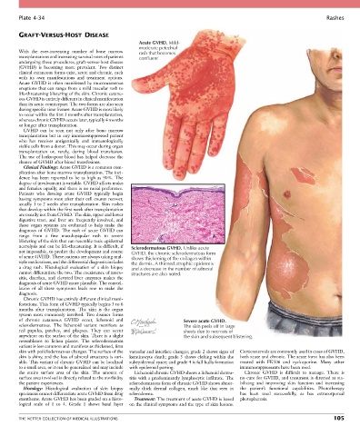
Plate 4-34                                                                                                           Rashes


        GRAFT-VERSUS-HOST DISEASE
                                                      Acute GVHD. Mild-
                                                      moderate petechial
        With  the  ever-increasing  number  of  bone  marrow   rash that becomes
        transplantations and increasing survival rates of patients   confluent
        undergoing these procedures, graft-versus-host disease
        (GVHD)  is  becoming  more  prevalent.  Two  distinct
        clinical cutaneous forms exist, acute and chronic, each
        with  its  own  manifestations  and  treatment  options.
        Acute  GVHD  is  often  manifested  by  mucocutaneous
        eruptions that can range from a mild macular rash to
        life-threatening blistering of the skin. Chronic cutane-
        ous GVHD is entirely different in clinical manifestation
        than its acute counterpart. The two forms are also seen
        during specific time frames: Acute GVHD is most likely
        to occur within the first 3 months after transplantation,
        whereas chronic GVHD occurs later, typically 4 months
        or longer after transplantation.
          GVHD  can  be  seen  not  only  after  bone  marrow
        transplantation  but  in  any  immunosuppressed  patient
        who  has  receives  antigenically  and  immunologically
        viable cells from a donor. This may occur during organ
        transplantation  or,  rarely,  during  blood  transfusion.
        The use of leuko-poor blood has helped decrease the
        chance of GVHD after blood transfusions.
          Clinical Findings: Acute GVHD is a common com-
        plication after bone marrow transplantation. The inci-
        dence  has  been  reported  to  be  as  high  as  90%.  The
        degree of involvement is variable. GVHD affects males
        and females equally, and there is no racial preference.
        Patients  who  develop  acute  GVHD  typically  begin
        having symptoms soon after their cell counts recover,
        usually 1 to 2 weeks after transplantation. Skin rashes
        that develop within the first week after transplantation
        are usually not from GVHD. The skin, upper and lower
        digestive  tract,  and  liver  are  frequently  involved,  and
        these  organ  systems  are  evaluated  to  help  make  the
        diagnosis  of  GVHD.  The  rash  of  acute  GVHD  can
        range  from  a  fine  maculopapular  rash  to  severe
        blistering of the skin that can resemble toxic epidermal
        necrolysis and can be life-threatening. It is difficult, if   Sclerodermatous GVHD. Unlike acute
        not impossible, to predict the development and course   GVHD, the chronic sclerodermatous form
        of acute GVHD. These patients are always taking mul-  shows thickening of the collagen within
        tiple medications, and the differential diagnosis includes   the dermis. A thinned atrophic epidermis
        a  drug  rash.  Histological  evaluation  of  a  skin  biopsy   and a decrease in the number of adnexal
        cannot differentiate the two. The coexistence of muco-  structures are also noted.
        sitis,  diarrhea,  and  elevated  liver  enzymes  makes  the
        diagnosis of acute GVHD more plausible. The constel-
        lation  of  all  these  symptoms  leads  one  to  make  the
        diagnosis.
          Chronic GVHD has entirely different clinical mani-
        festations. This form of GVHD typically begins 3 to 6
        months  after  transplantation.  The  skin  is  the  organ
        system  most  commonly  involved.  Two  distinct  forms
        of  chronic  cutaneous  GVHD  occur,  lichenoid  and           Severe acute GVHD.
        sclerodermatous.  The  lichenoid  variant  manifests  as       The skin peels off in large
        red  papules,  patches,  and  plaques.  They  can  occur       sheets due to necrosis of
        anywhere on the surface of the skin. There is a slight         the skin and subsequent blistering.
        resemblance  to  lichen  planus.  The  sclerodermatous
        variant is less common and manifests as thickened, firm
        skin with poikilodermatous changes. The surface of the   vacuolar and interface changes; grade 2 shows signs of   Corticosteroids are commonly used in cases of GVHD,
        skin is shiny, and the loss of adnexal structures is vari-  keratinocyte death; grade 3 shows clefting within the   both acute and chronic. The acute form has also been
        able. This variant of chronic GVHD can be localized   subepidermal space; and grade 4 is full bulla formation   treated  with  FK506  and  cyclosporine.  Many  other
        to a small area, or it can be generalized and may include   with epidermal parting.  immunosuppressants have been used.
        the  entire  surface  area  of  the  skin.  The  amount  of   Lichenoid chronic GVHD shows a lichenoid derma-  Chronic  GVHD  is  difficult  to  manage.  There  is
        surface area involved is directly related to the morbidity   titis with a predominantly lymphocytic infiltrate. The   no cure for GVHD, and treatment is directed at sta-
        the patient experiences.                  sclerodermatous form of chronic GVHD shows abnor-  bilizing  and  improving  skin  function  and  increasing
          Histology:  Histological  evaluation  of  skin  biopsy   mally  thick  dermal  collagen,  much  like  that  seen  in   the  patient’s  functional  capabilities.  Phototherapy
        specimens cannot differentiate acute GVHD from drug   scleroderma.                  has  been  used  successfully,  as  has  extracorporeal
        exanthems. Acute GVHD has been graded on a histo-  Treatment: The treatment of acute GVHD is based   photopheresis.
        logical  scale  of  1  to  4.  Grade  1  shows  basal  layer   on the clinical symptoms and the type of skin lesions.


        THE NETTER COLLECTION OF MEDICAL ILLUSTRATIONS                                                                          105