
Page 116 - The Netter Collection of Medical Illustrations - Integumentary System_ Volume 4 ( PDFDrive )
P. 116
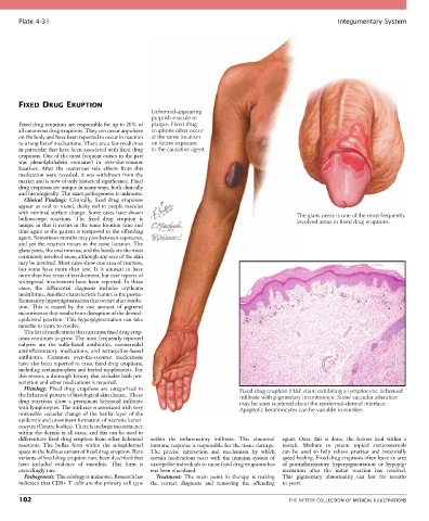
Plate 4-31                                                                                            Integumentary System


       FIXED DRUG ERUPTION
                                                 Lichenoid-appearing
                                                 purplish macule or
       Fixed drug eruptions are responsible for up to 20% of   plaque. Fixed drug
       all cutaneous drug eruptions. They can occur anywhere   eruptions often occur
       on the body and have been reported to occur in reaction   at the same location
       to a long list of medications. There are a few medicines   on future exposure
       in particular that have been associated with fixed drug   to the causative agent.
       eruptions. One of the most frequent causes in the past
       was  phenolphthalein  contained  in  over-the-counter
       laxatives.  After  the  numerous  side  effects  from  this
       medication were revealed, it was withdrawn from the
       market and is now of only historical significance. Fixed
       drug eruptions are unique in many ways, both clinically
       and histologically. The exact pathogenesis is unknown.
         Clinical  Findings:  Clinically,  fixed  drug  eruptions
       appear as oval to round, dusky red to purple macules
       with minimal surface change. Some cases have shown                                       The glans penis is one of the most frequently
       bullous-type  reactions.  The  fixed  drug  eruption  is                                 involved areas in fixed drug eruptions.
       unique in that it recurs in the same location time and
       time again as the patient is reexposed to the offending
       agent. Sometimes months may pass between exposures,
       and yet the reaction recurs in the same location. The
       glans penis, the oral mucosa, and the hands are the most
       commonly involved areas, although any area of the skin
       may be involved. Most cases show one area of reaction,
       but  some  have  more  than  one.  It  is  unusual  to  have
       more than five areas of involvement, but case reports of
       widespread involvement have been reported. In these
       cases,  the  differential  diagnosis  includes  erythema
       multiforme. Another characteristic feature is the postin-
       flammatory hyperpigmentation that occurs after resolu-
       tion.  This  is  caused  by  the  vast  amount  of  pigment
       incontinence that results from disruption of the dermal-
       epidermal  junction.  This  hyperpigmentation  can  take
       months to years to resolve.
         The list of medications that can cause fixed drug erup-
       tions continues to grow. The most frequently reported
       culprits  are  the  sulfa-based  antibiotics,  nonsteroidal
       antiinflammatory  medications,  and  tetracycline-based
       antibiotics.  Common  over-the-counter  medications
       have also been reported to cause fixed drug eruptions,
       including acetaminophen and herbal supplements. For
       this reason, a thorough history that includes both pre-
       scription and other medications is required.
         Histology:  Fixed  drug  eruptions  are  categorized  in            Fixed drug eruption (H&E stain) exhibiting a lymphocytic lichenoid
       the lichenoid pattern of histological skin disease. These             infiltrate with pigmentary incontinence. Some vacuolar alteration
       drug  reactions  show  a  prominent  lichenoid  infiltrate            may be seen scattered about the epidermal-dermal interface.
       with lymphocytes. The infiltrate is associated with very              Apoptotic keratinocytes can be variable in number.
       noticeable vacuolar change of the basilar layer of the
       epidermis and prominent formation of necrotic kerati-
       nocytes (Civatte bodies). There is melanin incontinence
       within the dermis in all cases, and this can be used to
       differentiate fixed drug eruption from other lichenoid   within  the  inflammatory  infiltrate.  This  abnormal   agent.  Once  this  is  done,  the  lesions  heal  within  a
       reactions.  The  bullae  form  within  the  subepidermal   immune response is responsible for the tissue damage.   month.  Medium  to  potent  topical  corticosteroids
       space in the bullous variant of fixed drug eruption. Rare   The  precise  interaction  and  mechanism  by  which   can  be  used  to  help  relieve  pruritus  and  potentially
       variants of fixed drug eruption have been described that   certain medications react with the immune system of   speed healing. Fixed drug eruptions often leave an area
       have  included  evidence  of  vasculitis.  This  form  is   susceptible individuals to cause fixed drug eruptions has   of  postinflammatory  hyperpigmentation  or  hypopig-
       exceedingly rare.                         not been elucidated.                      mentation  after  the  initial  reaction  has  resolved.
         Pathogenesis: The etiology is unknown. Research has   Treatment:  The  main  point  in  therapy  is  making   This  pigmentary  abnormality  can  last  for  months
       indicated that CD8+ T cells are the primary cell type   the  correct  diagnosis  and  removing  the  offending   to years.

       102                                                                                   THE NETTER COLLECTION OF MEDICAL ILLUSTRATIONS