
Page 136 - The Netter Collection of Medical Illustrations - Integumentary System_ Volume 4 ( PDFDrive )
P. 136
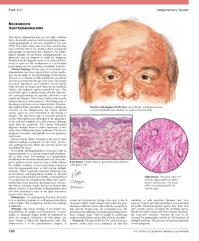
Plate 4-51                                                                                            Integumentary System


       NECROBIOTIC
       XANTHOGRANULOMA


       Necrobiotic xanthogranuloma is a rare skin condition
       that is frequently associated with an underlying mono-
       clonal gammopathy. It was first described in the early
       1980s. Since then, many cases have been reported that
       have confirmed this to be a distinct, albeit unusual and
       infrequently encountered, skin condition. The patho-
       logical  findings  of  necrobiotic  xanthogranuloma  are
       distinctive  and  are  required  to  make  the  diagnosis.
       Patients with this diagnosis need to be monitored rou-
       tinely to watch for the development of a monoclonal
       gammopathy and the possibility of multiple myeloma.
         Clinical Findings: So few cases of necrobiotic xan-
       thogranuloma have been reported that no firm conclu-
       sion can be made on the epidemiology of the disease.
       However, it is a disease of older adulthood, with almost
       all cases occurring after the age of 50 years. The lesions
       have been reported to occur anywhere on the human
       body, but they are found most often on the forehead,
       cheeks,  and  temporal  regions  around  the  eyes.  The
       periorbital region is almost always affected. Necrobi-
       otic  xanthogranulomas  are  typically  yellowish  to  red
       papules and plaques. There may be intervening atrophy
       between the areas of involvement. The leading edge of
       the plaques may have a red or violaceous hue. Occasion-
       ally, nodules form. Secondary ulceration is frequently   Patches and plaques on the face. Necrobiotic xanthogranuloma
       reported,  as  are  telangiectases  and  dilated  dermal   is most frequently encountered in a periocular location.
       vessels,  which  are  most  prominent  in  the  regions  of
       atrophy.  The  ulcerations  take  an  extended  period  to
       resolve. Most patients are distraught by the appearance
       of the rash and complain of a mild pruritus, although
       many  have  no  symptoms.  The  clinical  differential
       diagnosis includes forms of planar xanthomas. A skin
       biopsy helps differentiate these conditions. This disease
       progresses over time and typically does not spontane-
       ously remit.
         Patients almost always complain of dry eyes or have
       objective findings of proptosis. In rare cases, necrobi-
       otic  xanthogranuloma  affects  the  lacrimal  gland  and
       retrobulbar fat tissue.
         Necrobiotic xanthogranuloma is associated with an
       immunoglobulin  G-κ  (IgG:κ)  monoclonal  gammopa-
       thy  in  most  cases.  The  presence  of  a  gammopathy
       should make the clinician seek the advice of a hematolo-
       gist to perform a bone marrow biopsy to help evaluate   Low power. Diffuse dermal granulomatous infiltrate
       for multiple myeloma. A small percentage of patients     with giant cells
       with  this  gammopathy  have  or  will  develop  multiple
       myeloma. Other frequently abnormal laboratory tests
       in  necrobiotic  xanthogranuloma  include  an  elevated
       erythrocyte sedimentation rate (ESR), a decreased level                                      High power. The giant cells can
       of complement C4, and leukopenia. Many other abnor-                                          be best appreciated on higher-
       malities have been described, providing more evidence                                        power microscopy. The giant
       that this is a systemic disease and not an isolated skin                                     cells are predominantly the
       disease. Lesions of necrobiotic xanthogranuloma have                                         Touton type.
       also been described to occur in the upper respiratory
       system and in the heart.
         Pathogenesis: The pathogenesis has been theorized
       to be an antibody response to a self-antigen, most likely   unique and characteristic finding, when seen, is that of   condition,  so  only  anecdotal  therapies  have  been
       a form of lipid. This is unproven, and the exact etiology   cholesterol-filled, needle-shaped clefts within the gran-  reported. Topical and oral steroids have been somewhat
       is unknown.                               ulomatous infiltrate. Giant cells, both the foreign body   successful.  Chemotherapeutic  agents  have  been  used
         Histology:  The  biopsy  findings  of  necrobiotic  xan-  type  and  the  Touton  type,  are  commonly  seen.  The   with  variable  success,  including  the  alkylating  agents.
       thogranuloma are unique and characteristic. A punch   granulomatous infiltrate surrounds and envelops necro-  Results have been varied, with some patients experienc-
       biopsy  or  excisional  biopsy  should  be  performed  to   biotic collagen tissue. There is usually an underlying,   ing  long-term  remission.  Patients  all  need  to  be
       allow  for  adequate  evaluation.  On  first  glance,  the   predominantly lobular panniculitis without vasculitis.  screened for gammopathy and for the development of
       entire  dermis  is  filled  with  inflammatory  cells.  The   Treatment: Therapy is difficult. No randomized pro-  multiple myeloma. The presence of myeloma portends
       inflammation  is  in  the  granulomatous  category.  A   spective  studies  have  been  performed  on  this  rare   a worse prognosis.

       122                                                                                   THE NETTER COLLECTION OF MEDICAL ILLUSTRATIONS