
Page 139 - The Netter Collection of Medical Illustrations - Integumentary System_ Volume 4 ( PDFDrive )
P. 139
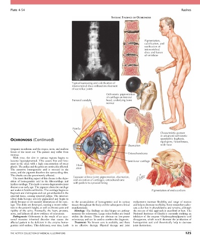
Plate 4-54                                                                                                           Rashes

                                                                             SYSTEMIC FINDINGS OF OCHRONOSIS


                                                                                                    Pigmentation,
                                                                                                    calcification, and
                                                                                                    ossification of
                                                                                                    intervertebral
                                                                                                    discs and fusion
                                                                                                    of vertebrae


                                                  Typical narrowing and calcification of
                                                  intervertebral discs without involvement
                                                  of sacroiliac joints

                                                                          Ochronotic pigmentation
                                                                          of cartilage on femoral
                                                  Femoral condyle         head; underlying bone
                                                                          normal


                                                   Patella
                                                                                                               Characteristic posture
                                                                                                               in advanced ochronotic
        OCHRONOSIS (Continued)                                                                                 spondylitis: kyphosis,
                                                                                                               rigid spine, flexed knees,
                                                                                         Eburnation            wide base
        tympanic membrane and the stapes, incus, and malleus
        bones  of  the  inner  ear.  The  patient  may  suffer  from                     Osteochondroma
        tinnitus.
          With  time,  the  skin  in  various  regions  begins  to
        become  hyperpigmented.  This  occurs  first  and  fore-                         Semilunar cartilage
        most in the areas with a high concentration of sweat   Ulcer-
        glands. The axillae and the groin are noticeably affected.   ation
        The  excessive  homogentisic  acid  is  secreted  in  the
        sweat, and the pigment discolors the surrounding skin.
        The cheeks are also prominently affected.
          The most disabling aspect of this disease is the depo-  Exposure of knee joint: pigmentation, eburnation,
        sition  of  homogentisic  acid  in  the  fibrocartilage  and   and ulceration of cartilages; osteochondroma
        hyaline cartilage. This leads to severe degenerative joint   with pedicle to synovial lining
        disease at an early age. The pigment alters the cartilage
        and makes it brittle and friable. The cartilage begins to                                        Pigmentation of endocardium
        fragment and disintegrate and can get embedded in the
        synovial tissue, causing synovial polyps. The interver-
        tebral  disks  become  severely  pigmented  and  begin  to
        calcify because of the massive destruction of the carti-  to  the  accumulation  of  homogentisic  acid  in  various   replacement  increase  flexibility  and  range  of  motion
        lage. The disks are destroyed, causing a severe reduc-  tissues throughout the body and the subsequent clinical   and help to decrease morbidity. Some researchers advo-
        tion in the patient’s height, as well as chronic pain and   manifestations.         cate a diet low in phenylalanine and tyrosine, although
        rigidity  of  the  spine.  Eventually,  the  heart,  prostate,   Histology: The findings on skin biopsy are pathog-  the success of this approach is anecdotal at best. The
        aorta, and kidneys all show evidence of ochronosis.  nomonic for ochronosis. Large ochre bodies are found   National Institutes of Health is currently studying an
          Pathogenesis:  Ochronosis  is  the  result  of  an  auto-  within  the  dermis.  These  are  obvious  on  low-power   inhibitor  of  the  enzyme  4-hydroxyphenylpyruvic  acid
        somal  recessive  inherited  disorder  that  causes  the   microscopy and can be used to confirm the diagnosis.  dioxygenase, which would decrease the production of
        affected patient to be deficient in the enzyme homo-  Treatment:  No  known  cure  is  available,  and  there   homogentisic  acid  and  theoretically  help  to  decrease
        gentisic acid oxidase. This deficiency, over time, leads   is  no  effective  therapy.  Physical  therapy  and  joint   joint destruction.


        THE NETTER COLLECTION OF MEDICAL ILLUSTRATIONS                                                                          125