
Page 141 - The Netter Collection of Medical Illustrations - Integumentary System_ Volume 4 ( PDFDrive )
P. 141
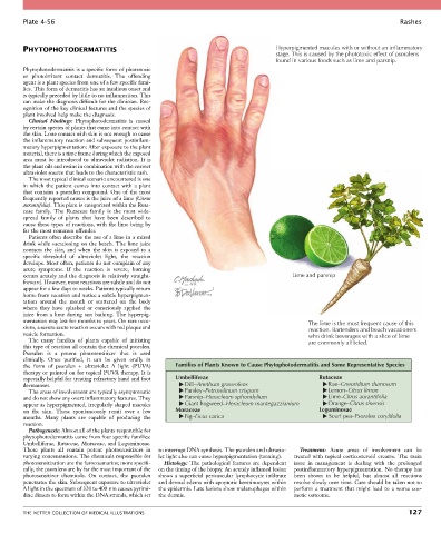
Plate 4-56                                                                                                           Rashes


        PHYTOPHOTODERMATITIS                                                           Hyperpigmented macules with or without an inflammatory
                                                                                       stage. This is caused by the phototoxic effect of psoralens
                                                                                       found in various foods such as lime and parsnip.
        Phytophotodermatitis is a specific form of phototoxic
        or  photoirritant  contact  dermatitis.  The  offending
        agent is a plant species from one of a few specific fami-
        lies. This form of dermatitis has an insidious onset and
        is typically preceded by little to no inflammation. This
        can make the diagnosis difficult for the clinician. Rec-
        ognition of the key clinical features and the species of
        plant involved help make the diagnosis.
          Clinical  Findings:  Phytophotodermatitis  is  caused
        by certain species of plants that come into contact with
        the skin. Lone contact with skin is not enough to cause
        the inflammatory reaction and subsequent postinflam-
        matory hyperpigmentation: After exposure to the plant
        material, there is a time frame during which the exposed
        area must be introduced to ultraviolet radiation. It is
        the plant oils and resins in combination with the correct
        ultraviolet source that leads to the characteristic rash.
          The most typical clinical scenario encountered is one
        in  which  the  patient  comes  into  contact  with  a  plant
        that contains a psoralen compound. One of the most
        frequently reported causes is the juice of a lime (Citrus
        aurantifolia). This plant is categorized within the Ruta-
        ceae  family.  The  Rutaceae  family  is  the  most  wide-
        spread  family  of  plants  that  have  been  described  to
        cause these types of reactions, with the lime being by
        far the most common offender.
          Patients often describe the use of a lime in a mixed
        drink while vacationing on the beach. The lime juice
        contacts  the  skin,  and  when  the  skin  is  exposed  to  a
        specific  threshold  of  ultraviolet  light,  the  reaction
        develops. Most often, patients do not complain of any
        acute  symptoms.  If  the  reaction  is  severe,  burning
        occurs acutely and the diagnosis is relatively straight-                            Lime and parsnip
        forward. However, most reactions are subtle and do not
        appear for a few days to weeks. Patients typically return
        home from vacation and notice a subtle hyperpigmen-
        tation  around  the  mouth  or  scattered  on  the  body
        where  they  have  splashed  or  consciously  applied  the
        juice from a lime during sun bathing. The hyperpig-
        mentation may last for months to years. On rare occa-                                    The lime is the most frequent cause of this
        sions, a severe acute reaction occurs with red plaque and                                reaction. Bartenders and beach vacationers
        vesicle formation.                                                                       who drink beverages with a slice of lime
          The  many  families  of  plants  capable  of  initiating                               are commonly afflicted.
        this type of reaction all contain the chemical psoralen.
        Psoralen  is  a  potent  photosensitizer  that  is  used
        clinically.  Once  purified,  it  can  be  given  orally  in
        the  form  of  psoralen  +  ultraviolet  A  light  (PUVA)   Families of Plants Known to Cause Phytophotodermatitis and Some Representative Species
        therapy or painted on for topical PUVA therapy. It is
        especially helpful for treating refractory hand and foot   Umbelliferae                     Rutaceae
        dermatoses.                                       Dill–Anethum graveolens                      Rue–Cneoridium dumosum
          The areas of involvement are typically asymptomatic   Parsley–Petroselinum crispum           Lemon–Citrus limon
        and do not show any overt inflammatory features. They   Parsnip–Heracleum sphondylium          Lime–Citrus aurantifolia
        appear as hyperpigmented, irregularly shaped macules   Giant hogweed–Heracleum mantegazzianium  Orange–Citrus sinensis
        on  the  skin.  These  spontaneously  remit  over  a  few   Moraceae                        Leguminosae
        months.  Many  plants  are  capable  of  producing  the   Fig–Ficus carica                     Scurf pea–Psoralea corylifolia
        reaction.
          Pathogenesis: Almost all of the plants responsible for
        phytophotodermatitis come from four specific families:
        Umbelliferae, Rutaceae, Moraceae, and Leguminosae.
        These  plants  all  contain  potent  photosensitizers  in   to interrupt DNA synthesis. The psoralen and ultravio-  Treatment:  Acute  areas  of  involvement  can  be
        varying concentrations. The chemicals responsible for   let light also can cause hyperpigmentation (tanning).  treated  with  topical  corticosteroid  creams.  The  main
        photosensitization are the furocoumarins; more specifi-  Histology: The pathological features are dependent   issue  in  management  is  dealing  with  the  prolonged
        cally, the psoralens are by far the most important of the   on the timing of the biopsy. An acutely inflamed lesion   postinflammatory hyperpigmentation. No therapy has
        photosensitizer  chemicals.  On  contact,  the  psoralen   shows  a  superficial  perivascular  lymphocytic  infiltrate   been  shown  to  be  helpful,  but  almost  all  reactions
        penetrates the skin. Subsequent exposure to ultraviolet   and dermal edema with apoptotic keratinocytes within   resolve slowly over time. Care should be taken not to
        A light in the spectrum of 320 to 400 nm causes pyrimi-  the epidermis. Late lesions show melanophages within   perform  a  treatment  that  might  lead  to  a  worse  cos-
        dine dimers to form within the DNA strands, which act   the dermis.                 metic outcome.


        THE NETTER COLLECTION OF MEDICAL ILLUSTRATIONS                                                                          127