
Page 144 - The Netter Collection of Medical Illustrations - Integumentary System_ Volume 4 ( PDFDrive )
P. 144
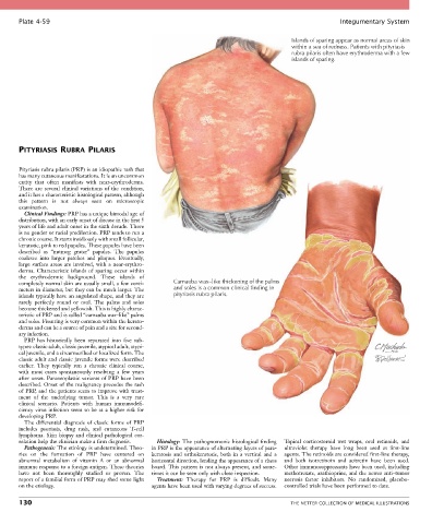
Plate 4-59                                                                                            Integumentary System

                                                                                             Islands of sparing appear as normal areas of skin
                                                                                             within a sea of redness. Patients with pityriasis
                                                                                             rubra pilaris often have erythroderma with a few
                                                                                             islands of sparing.


       PITYRIASIS RUBRA PILARIS


       Pityriasis rubra pilaris (PRP) is an idiopathic rash that
       has many cutaneous manifestations. It is an uncommon
       entity  that  often  manifests  with  near-erythroderma.
       There  are  several  clinical  variations  of  the  condition,
       and it has a characteristic histological pattern, although
       this  pattern  is  not  always  seen  on  microscopic
       examination.
         Clinical Findings: PRP has a unique bimodal age of
       distribution, with an early onset of disease in the first 5
       years of life and adult onset in the sixth decade. There
       is no gender or racial predilection. PRP tends to run a
       chronic course. It starts insidiously with small follicular,
       keratotic, pink to red papules. These papules have been
       described  as  “nutmeg  grater”  papules.  The  papules
       coalesce  into  larger  patches  and  plaques.  Eventually,
       large  surface  areas  are  involved,  with  a  near-erythro-
       derma.  Characteristic  islands  of  sparing  occur  within
       the  erythrodermic  background.  These  islands  of
       completely normal skin are usually small, a few centi-  Carnauba wax–like thickening of the palms
       meters in diameter, but they can be much larger. The   and soles is a common clinical finding in
       islands typically have an angulated shape, and they are   pityriasis rubra pilaris.
       rarely  perfectly  round  or  oval.  The  palms  and  soles
       become thickened and yellowish. This is highly charac-
       teristic of PRP and is called “carnauba wax–like” palms
       and soles. Fissuring is very common within the kerato-
       derma and can be a source of pain and a site for second-
       ary infection.
         PRP  has  historically  been  separated  into  five  sub-
       types: classic adult, classic juvenile, atypical adult, atypi-
       cal juvenile, and a circumscribed or localized form. The
       classic adult and classic juvenile forms were described
       earlier.  They  typically  run  a  chronic  clinical  course,
       with  most  cases  spontaneously  resolving  a  few  years
       after onset. Paraneoplastic variants of PRP have been
       described. Onset of the malignancy precedes the rash
       of PRP, and the patients seem to improve with treat-
       ment  of  the  underlying  tumor.  This  is  a  very  rare
       clinical  scenario.  Patients  with  human  immunodefi-
       ciency virus infection seem to be at a higher risk for
       developing PRP.
         The  differential  diagnosis  of  classic  forms  of  PRP
       includes  psoriasis,  drug  rash,  and  cutaneous  T-cell
       lymphoma.  Skin  biopsy  and  clinical  pathological  cor-
       relation help the clinician make a firm diagnosis.  Histology:  The  pathognomonic  histological  finding   Topical  corticosteroid  wet  wraps,  oral  retinoids,  and
         Pathogenesis: The etiology is undetermined. Theo-  in PRP is the appearance of alternating layers of para-  ultraviolet  therapy  have  long  been  used  as  first-line
       ries  on  the  formation  of  PRP  have  centered  on     keratosis  and  orthokeratosis,  both  in  a  vertical  and  a   agents. The retinoids are considered first-line therapy,
       abnormal  metabolism  of  vitamin  A  or  an  abnormal   horizontal direction, lending the appearance of a chess   and  both  isotretinoin  and  acitretin  have  been  used.
       immune response to a foreign antigen. These theories   board. This pattern is not always present, and some-  Other immunosuppressants have been used, including
       have  not  been  thoroughly  studied  or  proven.  The   times it can be seen only with close inspection.  methotrexate, azathioprine, and the newer anti–tumor
       report of a familial form of PRP may shed some light   Treatment:  Therapy  for  PRP  is  difficult.  Many   necrosis  factor  inhibitors.  No  randomized,  placebo-
       on the etiology.                          agents have been used with varying degrees of success.   controlled trials have been performed to date.

       130                                                                                   THE NETTER COLLECTION OF MEDICAL ILLUSTRATIONS