
Page 142 - The Netter Collection of Medical Illustrations - Integumentary System_ Volume 4 ( PDFDrive )
P. 142
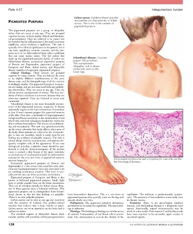
Plate 4-57                                                                                            Integumentary System

                                                      Lichen aureus. Golden-colored macules
       PIGMENTED PURPURA                              and patches are characteristic of lichen
                                                      aureus. This is one of the variants of
                                                      pigmented purpura.
       The  pigmented  purpuras  are  a  group  of  idiopathic
       rashes  that  can  occur  at  any  age.  They  are  grouped
       together because of their similar clinical and histologi-
       cal presentations. They are believed to be caused not
       by vasculitis but by inflammation of the small cutaneous
       capillaries,  which  produces  a  capillaritis.  The  rash  is
       typically of no clinical significance to the patient, but it
       can  cause  significant  cosmetic  concern,  and  the  pur-
       puras need to be differentiated from other conditions
       that  can  cause  similar  rashes.  The  five  rashes  that
       make up the pigmented purpuric family of rashes are   Schamberg’s disease. Cayenne
       Schamberg’s  disease,  eczematous  pigmented  purpura     pepper–like petechiae.
       of  Doucas  and  Kapetanakis,  pigmented  purpura  of   This asymptomatic
       Gougerot  and  Blum,  lichen  aureus,  and  Majocchi’s   idiopathic rash is almost
       disease (annular telangiectatic pigmented purpura).  exclusively seen on the
         Clinical  Findings:  These  entities  are  grouped   lower legs.
       together for many reasons. They are believed by some
       to  be  slightly  different  manifestations  of  the  same
       disease state, and the histopathology of all the variants
       is strikingly similar. The pigmented purpuric dermato-
       ses are benign and are not associated with any underly-
       ing abnormality. They can occur at any age. They are
       almost entirely asymptomatic in nature. The true inci-
       dence of these conditions is unknown, because they are
       often  not  reported.  They  are  believed  to  occur  very
       commonly.
         Schamberg’s disease is the most frequently encoun-
       tered  of  the  pigmented  purpuric  eruptions.  It  almost
       universally begins on the lower extremities. It manifests
       as tiny (1-mm) cayenne pepper–like petechial macules
       of the skin. Over time, a brownish-red hyperpigmented
       background forms secondary to the extravasation of red
       blood cells and their subsequent breakdown within the
       skin to release hemosiderin. The lesions are nonblanch-
       ing and nonpalpable. The rash may spread proximally
       up the lower extremity but rarely affects other areas of
       the body. Most patients are referred to the dermatolo-
       gist to rule out vasculitis, which is easily done by not
       finding any evidence of palpable purpura. The rash is
       almost always entirely asymptomatic, and patients fre-
       quently complain only of the appearance. If one sees
       widespread  petechia,  a  platelet  count  should  be  per-
       formed  to  look  for  thrombocytopenia.  If  the  platelet
       count is normal, a skin biopsy of the upper extremity
       or truncal area of involvement should be performed to
       evaluate for the very rare form of pigmented purpuric                      Extravasated erythrocytes and a lymphocytic vasculitis are the
       mycosis fungoides.                                                         key histological features.
         Eczematoid  pigmented  purpura  of  Doucas  and
       Kapetanakis is a rare variant that manifests with pete-
       chiae and hyperpigmentation but is also associated with
       an overlying eczematous eruption. This form is typi-
       cally pruritic and can show secondary excoriations.
         Pigmented  purpura  of  Gougerot  and  Blum  is  also
       known  as  lichenoid  pigmented  purpura.  Small,  light
       pink to purple papules form on the lower extremities.
       They can be mistaken initially for lichen planus. Biop-
       sies of these papules show a lichenoid infiltrate. This
       pigmented purpura can be distinguished from Scham-
       berg’s  disease  in  that  the  skin  findings  are  palpable.   from  hemosiderin  deposition.  This  is  a  rare  form  of   capillaritis.  The  infiltrate  is  predominantly  lympho-
       There is no true palpable purpura.        pigmented purpura that typically starts on the legs and   cytic. The presence of hemosiderin is more easily seen
         Lichen aureus can be seen at any age and manifests   spreads slowly over time.    in chronic lesions.
       with  the  presence  of  multiple  tiny,  golden-colored   Pathogenesis:  The  pigmented  purpuric  dermatoses   Treatment:  There  is  no  agreed-upon  standard
       macules  that  coalesce  into  a  large  macule  or  patch.   are believed to be caused by capillaritis. The exact etiol-  therapy, and withholding therapy is a frequently used
       Lichen aureus can occur anywhere on the body and is   ogy is unknown.               option.  Anecdotally,  topical  corticosteroids  may  be
       solitary in nature.                         Histology: The histological findings are similar across   tried for a few weeks. Oral vitamin C and bioflavonoids
         The  involved  regions  in  Majocchi’s  disease  show   all variants. Extravasation of red blood cells is promi-  have  been  reported  to  be  successful,  again  mostly  in
       annular patches with petechiae and hyperpigmentation   nent.  The  extravasation  is  seen  in  the  vicinity  of  the   anecdotal reports.

       128                                                                                   THE NETTER COLLECTION OF MEDICAL ILLUSTRATIONS