
Page 143 - The Netter Collection of Medical Illustrations - Integumentary System_ Volume 4 ( PDFDrive )
P. 143
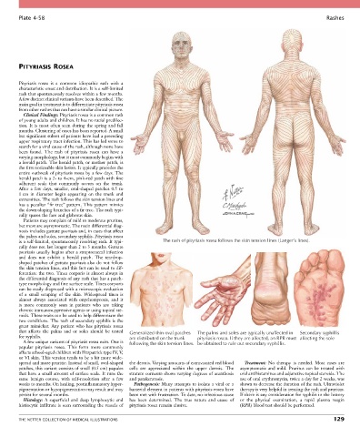
Plate 4-58                                                                                                           Rashes


        PITYRIASIS ROSEA


        Pityriasis  rosea  is  a  common  idiopathic  rash  with  a
        characteristic onset and distribution. It is a self-limited
        rash that spontaneously resolves within a few months.
        A few distinct clinical variants have been described. The
        main goal in treatment is to differentiate pityriasis rosea
        from other rashes that can have a similar clinical picture.
          Clinical Findings: Pityriasis rosea is a common rash
        of young adults and children. It has no racial predilec-
        tion.  It  is  most  often  seen  during  the  spring  and  fall
        months. Clustering of cases has been reported. A small
        but significant subset of patients have had a preceding
        upper respiratory tract infection. This has led some to
        search for a viral cause of the rash, although none have
        been  found.  The  rash  of  pityriasis  rosea  can  have  a
        varying morphology, but it most commonly begins with
        a herald patch. The herald patch, or mother patch, is
        the first noticeable skin lesion. It typically precedes the
        entire outbreak of pityriasis rosea by a few days. The
        herald patch is a 2- to 4-cm, pink-red patch with fine
        adherent  scale  that  commonly  occurs  on  the  trunk.
        After  a  few  days,  smaller,  oval-shaped  patches  0.5  to
        1 cm  in  diameter  begin  appearing  on  the  trunk  and
        extremities. The rash follows the skin tension lines and
        has a peculiar “fir tree” pattern. This pattern mimics
        the down-sloping branches of a fir tree. The rash typi-
        cally spares the face and glabrous skin.
          Patients may complain of mild to moderate pruritus,
        but most are asymptomatic. The main differential diag-
        nosis includes guttate psoriasis and, in cases that affect
        the palms and soles, secondary syphilis. Pityriasis rosea
        is a self-limited, spontaneously resolving rash. It typi-  The rash of pityriasis rosea follows the skin tension lines (Langer’s lines).
        cally does not last longer than 2 to 3 months. Guttate
        psoriasis usually begins after a streptococcal infection
        and  does  not  exhibit  a  herald  patch.  The  teardrop-
        shaped patches of guttate psoriasis also do not follow
        the skin tension lines, and this fact can be used to dif-
        ferentiate the two. Tinea corporis is almost always in
        the differential diagnosis of any rash that has a patch-
        type morphology and fine surface scale. Tinea corporis
        can be easily diagnosed with a microscopic evaluation
        of  a  small  scraping  of  the  skin.  Widespread  tinea  is
        almost  always  associated  with  onychomycosis,  and  it
        is  more  commonly  seen  in  patients  who  are  taking
        chronic immunosuppressive agents or using topical ste-
        roids. These traits can be used to help differentiate the
        two conditions. The rash of secondary syphilis is the
        great  mimicker.  Any  patient  who  has  pityriasis  rosea
        that  affects  the  palms  and  or  soles  should  be  tested     Generalized thin oval patches  The palms and soles are typically unaffected in  Secondary syphillis
        for syphilis.                             are distributed on the trunk  pityriasis rosea. If they are affected, an RPR must affecting the sole
          A few unique variants of pityriasis rosea exist. One is   following the skin tension lines. be obtained to rule out secondary syphillis.
        papular  pityriasis  rosea.  This  form  more  commonly
        affects school-aged children with Fitzpatrick type IV, V,
        or VI skin. This version tends to be a bit more wide-
        spread and more pruritic. Instead of small, oval-shaped   the dermis. Varying amounts of extravasated red blood   Treatment:  No  therapy  is  needed.  Most  cases  are
        patches, this variant consists of small (0.5 cm) papules   cells  are  appreciated  within  the  upper  dermis.  The   asymptomatic  and  mild.  Pruritus  can  be  treated  with
        that have a small amount of surface scale. It runs the   stratum corneum shows varying degrees of acanthosis   oral antihistamines and adjunctive topical steroids. The
        same  benign  course,  with  self-resolution  after  a  few   and parakeratosis.    use of oral erythromycin, twice a day for 2 weeks, was
        weeks to months. On healing, postinflammatory hyper-  Pathogenesis: Many attempts to isolate a viral or a   shown to decrease the duration of the rash. Ultraviolet
        pigmentation or hypopigmentation may result and may   bacterial element in patients with pityriasis rosea have   therapy is very helpful in treating the rash and pruritus.
        persist for several months.               been met with frustration. To date, no infectious cause   If there is any consideration for syphilis in the history
          Histology:  A  superficial  and  deep  lymphocytic  and   has  been  determined.  The  true  nature  and  cause  of   or  the  physical  examination,  a  rapid  plasma  reagin
        histiocytic infiltrate is seen surrounding the vessels of   pityriasis rosea remain elusive.  (RPR) blood test should be performed.


        THE NETTER COLLECTION OF MEDICAL ILLUSTRATIONS                                                                          129