
Page 159 - The Netter Collection of Medical Illustrations - Integumentary System_ Volume 4 ( PDFDrive )
P. 159
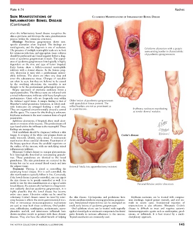
Plate 4-74                                                                                                           Rashes
        SKIN MANIFESTATIONS OF                                    CUTANEOUS MANIFESTATIONS OF INFLAMMATORY BOWEL DISEASE
        INFLAMMATORY BOWEL DISEASE
        (Continued)


        when  the  inflammatory  bowel  disease  re cognizes  the
        skin as gut tissue and develops the same granulomatous
        process within the cutaneous structures.
          Histology:  Pyoderma  gangrenosum  shows  non-
        specific  ulceration  when  biopsied.  The  findings  are
        nondiagnostic,  and  the  diagnosis  is  one  of  exclusion.                                   Cribiform ulceration with a purple
        The presence of multiple neutrophils leads one to look                                         surrounding border is characteristic
        for cutaneous infection, and appropriate tissue cultures                                       of pyoderma grangrenosum.
        should be performed and found negative before a diag-
        nosis of pyoderma gangrenosum is made. The appear-
        ance of pyoderma gangrenosum histologically is highly
        dependent  on  the  time  and  type  of  lesion  biopsied.
        Early  lesions  show  a  follicle-centered  neutrophilic
        infiltrate  with  a  dermal  abscess.  As  the  lesions  prog-
        ress,  ulceration  is  seen  with  a  predominant  neutro-
        philic  infiltrate.  The  ulcers  are  often  very  deep  and
        enter  the  subcutaneous  tissue.  Changes  of  vasculitis
        can often be seen, but they are believed to be caused
        by  the  overlying  ulceration;  the  vasculitis  is  not
        thought to be the predominant pathological process.
          Biopsy  specimens  of  erythema  nodosum  shows  a
        septal panniculitis. The fibrous septa are inflamed with
        a mixed inflammatory infiltrate with heavy lymphocyte
        predominance.  Giant  cells  are  frequently  seen  within
        the widened septal tissue. A unique finding is that of   Older lesion of pyoderma gangrenosum
        Miescher’s radial granuloma formation, in which mul-  with granulation tissue present. The
        tiple  histiocytes  are  arranged  flanking  a  small  area.   rolled borders are not as prominent as
        They are organized circumferentially around a central   in acute lesions.              Erythema nodosum manifesting
        slit-like space. The reason for this finding is unknown.                               as tender dermal nodules
        Erythema nodosum is the most common form of septal
        panniculitis.
          Aphthous ulcerations, if biopsied, show small ulcer-
        ations or erosions of the mucosa. The predominant cell
        type found within the infiltrate is the neutrophil. These
        findings are nonspecific.
          Oral candidiasis should be diagnosed without a skin
        biopsy. A scraping of the white oral plaques shows an                          Crohn’s disease
        easily  removed,  whitish,  sticky  tissue.  A  microscopic
        examination shows candidal elements. Examination of
        the biopsy specimen shows the candidal organisms on
        the  surface  of  the  mucosa,  with  an  underlying  mixed
        inflammatory infiltrate.
          Metastatic Crohn’s disease is a unique phenomenon.
        It is histologically described as noncaseating granulo-
        mas.  These  granulomas  are  identical  to  the  bowel
        granulomas. The skin granulomas are centered in the
        dermis but can be seen around blood vessels and into
        the adipose tissue.                       External fistula (via appendectomy incision)
          Treatment:  Therapy  is  aimed  at  controlling  the
        underlying  bowel  disease.  If  it  is  well  controlled,  the
        skin manifestations typically follow in line. Conversely,
        if the bowel disease is poorly controlled, one can expect
        the  skin  disease  to  be  poorly  controlled  as  well.  It  is
        useful to use the skin manifestations as a sign of active                                 Perianal fistulae and/or abscesses
        bowel disease. If a patient who has been in a long remis-
        sion  suddenly  develops  pyoderma  gangrenosum,  it  is
        highly  plausible  that  the  bowel  disease  has  become
        active  once  more.  Ulcerative  colitis  can  be  cured  by
        colectomy. Crohn’s disease cannot be cured by colec-  the  skin  disease.  Cyclosporine  and  prednisone  have   Erythema  nodosum  can  be  treated  with  compres-
        tomy because it affects the entire gastrointestinal tract.   shown excellent results in treating pyoderma gangreno-  sion  stockings,  topical  potent  steroids,  and  oral  ste-
        Oral  or  intravenous  immunosuppressive  medications   sum. Intralesional triamcinolone can be attempted on   roids  in  severe  cases.  Intralesional  injection  of
        are  used  to  treat  both  these  conditions.  Oral  predni-  small, early lesions of pyoderma gangrenosum.  triamcinolone  is  also  effective.  Metastatic  Crohn’s
        sone, sulfasalazine, azathioprine, methotrexate, myco-  Oral  aphthous  ulcers  can  be  treated  with  topically   disease  is  difficult  to  treat  and  requires  systemic
        phenolate  mofetil,  and  intravenous  infliximab  have   applied steroid gels or ointments compounded in dental   immunosuppressive agents such as azathioprine, pred-
        shown excellent results in patients with these chronic   paste  formula  to  increase  adherence  to  the  mucosa.   nisone,  or  infliximab.  It  is  best  treated  by  a  multi-
        diseases. They also have the added benefit of helping   Topical anesthetics are commonly used.  disciplinary  approach.


        THE NETTER COLLECTION OF MEDICAL ILLUSTRATIONS                                                                          145