
Page 160 - The Netter Collection of Medical Illustrations - Integumentary System_ Volume 4 ( PDFDrive )
P. 160
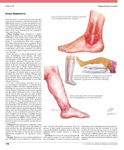
Plate 4-75                                                                                            Integumentary System


       STASIS DERMATITIS
                                                          Stasis dermatitis of the lower extremity appearing
                                                          as a hyperpigmented brown-red patch
       Stasis dermatitis is a common chronic dermatosis that
       is seen almost exclusively on the lower extremities. The
       inflammation can lead to chronic discoloration, ulcer-
       ation, and infection. Underlying systemic disease such
       as congestive heart failure and renal failure can predis-
       pose to stasis dermatitis. Any condition that can cause
       edema  of  the  lower  extremities  has  the  potential  to
       cause stasis dermatitis.
         Clinical  Findings:  Stasis  dermatitis  is  a  chronic
       inflammatory  skin  disease  that  indicates  underlying
       insufficiency  of  the  venous  return  system.  It  is  most
       commonly seen in the older population, and there is no
       gender  or  racial  predilection.  Most  often,  congestive
       heart failure is the associated disease causing the edema.
       Many other conditions of venous insufficiency can also
       be causative, including varicose veins and postsurgical
       complications,  such  as  after  a  saphenous  vein  harvest
       for coronary artery bypass surgery or an inguinal lymph
       node dissection.
         Stasis  dermatitis  is  a  skin  manifestation  of  a  wide
       range  of  underlying  venous  diseases.  The  lower
       extremities  account  for  more  than  99%  of  cases  of
       stasis dermatitis, and the diagnosis in other areas of the
       body  should  be  questioned.  The  legs  tend  to  have  a
       range of edema, from the very mild amount that accu-
       mulates at the end of a long day of standing to severe
       chronic  edema  that  is  always  present.  Red-brown
       patches,  some  with  a  light  yellow  discoloration,  typi-
       cally begin around the medial malleolus. As the condi-
       tion  progresses,  the  patches  begin  to  spread  and  can
       encompass the entire lower extremity, although much
       more  commonly  they  are  found  at  knee  level  or  just                Compression dressings or stockings are one of the best
       below knee level. There can be complete confluence of                      ways to keep fluid from accumulating in the lower
       the dermatitis around the affected limb, or it can affect                  extremities. Leg elevation is a mainstay of therapy.
       only part of the leg.
         The  rash  is  almost  always  symmetric,  and  it  is
       not  uncommonly  misdiagnosed  as  bilateral  lower
       extremity cellulitis. The rash is typically pruritic, and
       the itching can be so severe as to cause excoriations and
       small ulcerations. Depending on the severity, weeping
       vesicular patches and plaques can form. A rare bulla can
       also  be  seen  in  some  cases,  and  one  must  consider
       bullous pemphigoid in the differential diagnosis. Vari-                Venous stasis ulcerations are one complication
       cose veins are often present on examination, or there                  from long-standing or severe stasis dermatitis.
       may  be  a  history  of  bypass  surgery.  If  left  untreated,
       venous stasis can lead to venous ulcerations, which have
       been  described  as  slightly  painful  ulcerations  on  the
       lateral malleolus. The ulcerations can occur anywhere
       on the leg and in some cases are very tender. Peripheral
       pulses are intact, and this physical examination finding
       helps to rule out arterial insufficiency. If the ulcerations   Ulcer
       and edema are not controlled, the ulcerations will con-
       tinue to expand and can become secondarily infected;
       if they become deep enough, they can lead to underly-
       ing  osteomyelitis  or  cellulitis.  These  neglected  cases
       can end in  loss  of  the affected  portion  of  the limb  if
       medical therapies do not successfully clear the infection
       and ulcerations.
         Pathogenesis: Increased pressure within the venous
       system  of  the  lower  extremity  causes  extravasation  of   made  clinically.  Histological  examination  shows  an   Depending on the underlying reason for the stasis der-
       serum and blood into the surrounding dermis and sub-  increase  in  small  vessels,  extravasation  of  red  blood   matitis,  this  may  or  may  not  be  possible.  If  it  is  not
       cutaneous tissue. As the edema in the lower extremity   cells,  and  hemosiderin  deposition  in  the  dermis.  The   possible, the mainstay of therapy is the use of compres-
       worsens,  the  skin  begins  to  develop  signs  of  chronic   epidermis  shows  varying  amounts  of  spongiotic   sion stockings or wraps. However, the compliance rate
       inflammation  mediated  by  the  abnormal  location     dermatitis.                 is low because of difficulty putting them on and discom-
       of fluid.                                   Treatment: The rash can be treated symptomatically   fort. Those patients who are able to use the compres-
         Histology:  Biopsies  are  not  routinely  performed  in   with topical corticosteroids and emollients. The main   sion gear and topical corticosteroids usually have a good
       stasis  dermatitis,  and  the  diagnosis  is  almost  always   goal  of  therapy  is  to  restore  the  proper  venous  flow.   prognosis.

       146                                                                                   THE NETTER COLLECTION OF MEDICAL ILLUSTRATIONS