
Page 155 - The Netter Collection of Medical Illustrations - Integumentary System_ Volume 4 ( PDFDrive )
P. 155
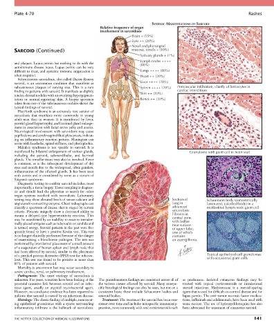
Plate 4-70                                                                                                           Rashes

                                                                             SYSTEMIC MANIFESTATIONS OF SARCOID
                                                   Relative frequency of organ
                                                   involvement in sarcoidosis
                                                                   Brain + (15%)
                                                                   Eyes ++ (20%)
                                                                   Nasal and pharyngeal
        SARCOID (Continued)                                        mucosa, tonsils + (10%)
                                                                       Salivary glands + (1%)
                                                                       Lymph nodes ++++
        and plaques. Lupus pernio has nothing to do with the           (80%)
        autoimmune disease lupus. Lupus pernio can be very
        difficult to treat, and systemic immune suppression is         Lungs ++++ (80%)
        often required.                                                Heart ++ (20%)
          Subcutaneous sarcoidosis, also called Darier-Roussy
        sarcoid,  is  an  uncommon  condition  that  manifests  as     Liver ++++ (70%)
        subcutaneous  plaques  of  varying  size.  This  is  a  rare   Spleen ++++ (70%)   Perivascular infiltration, chiefly of histiocytes in
        finding in patients with sarcoid. It manifests as slightly     Skin ++ (30%)     cardiac interstitium
        tender, dermal nodules with an overlying hyperpigmen-
        tation  or  normal-appearing  skin.  A  biopsy  specimen       Bones ++ (30%)
        taken from one of the subcutaneous nodules shows the
        typical findings of sarcoid.
          Heerfordt syndrome is an extremely rare version of
        sarcoidosis  that  manifests  more  commonly  in  young
        adult  men  than  in  women.  It  is  manifested  by  fever,
        parotid gland hypertrophy, and lacrimal gland enlarge-
        ment in association with facial nerve palsy and uveitis.
        Neurological  involvement  with  sarcoidosis  may  cause
        papilledema and cerebrospinal fluid pleocytosis, indicat-
        ing an inflammatory reaction pattern. Meningism can
        occur with headache, spinal stiffness, and photophobia.
          Mikulicz  syndrome  is  not  specific  to  sarcoid.  It  is
        manifested by bilateral enlargement of various glands,                                  Granuloma with giant cell in heart wall
        including  the  parotid,  submandibular,  and  lacrimal
        glands. The tonsillar tissue may also be involved. Fever
        is  common,  as  is  the  subsequent  development  of  dry
        eyes and mouth due to the widespread, often painless,
        inflammation of the affected glands. It has been seen
        with uveitis and is considered by some as a variant of
        Sjögren’s syndrome.
          Diagnostic testing to confirm sarcoid includes, most
        importantly, a tissue biopsy. Tissue sampling is diagnos-
        tic  and  should  lead  the  physician  to  search  for  other
        organ  systems  involved  with  sarcoidosis.  Laboratory
        testing may show elevated levels of serum calcium and                          Sectioned        Schaumann body (concentrically
        angiotensin-converting enzyme. Chest radiographs can                           lung in          laminated, calcified body) in a
        identify a spectrum of disease that is staged by certain                       advanced         mediastinal lymph node giant cell
        criteria. Patients uniquely show a decreased ability to                        sarcoidosis.
        mount  a  delayed-type  hypersensitivity  reaction.  This                      Fibrosis in
        may be manifested by an inability to react to intrader-                        central zone
                                                                                       with bullae
        mally placed antigens such as tuberculin or candida and                        near surface
        is termed anergy. Sarcoid patients in the past were fre-                       of upper lobe,
        quently found to have a positive Kveim test. This test                         one of which
        is no longer clinically performed because of the danger                        contains
        of transmitting a bloodborne pathogen. The test was                            an aspergilloma.
        performed by interdermal placement of a small amount
        of a suspension of human spleen and lymph node that
        had been affected by sarcoid, similar to the placement
        of a purified protein derivative (PPD) test for tubercu-                                        Typical epithelioid cell granulomas
        losis. This test was found to be positive in more than                                          with occasional giant cells
        85% of patients with sarcoid.
          Mortality is uncommon but may occur secondary to
        severe cardiac, renal, or pulmonary involvement.
          Pathogenesis:  The  exact  etiology  of  sarcoidosis  is
        unknown. For years, scientists have been looking at the   The granulomatous findings are consistent across all of   as  prednisone.  Isolated  cutaneous  findings  may  be
        potential causative link between sarcoid and an infec-  the various tissues affected by sarcoid. Many nonspe-  treated  with  topical  corticosteroids  or  intralesional
        tious  agent,  usually  an  atypical  mycobacterial  agent.   cific histological findings can also be seen, but not on a   steroid  injections.  Methotrexate  is  a  steroid-sparing
        However, no conclusive evidence has been reported to   consistent basis; these include Schaumann bodies and   agent that is used for difficult-to-control disease and for
        indicate that sarcoid is caused by an infectious disease.  asteroid bodies.         lupus pernio. The anti–tumor necrosis factor medica-
          Histology: The classic finding of multiple, noncaseat-  Treatment: The treatment for sarcoid has been con-  tions, infliximab and adalimumab, have been used with
        ing epithelioid granulomas with a sparse surrounding   sistent over time and includes nonspecific immunosup-  some success. The use of hydroxychloroquine has also
        inflammatory  infiltrate  is  the  hallmark  of  sarcoidosis.   pression, most commonly with oral corticosteroids such   been advocated for treatment of cutaneous sarcoid.


        THE NETTER COLLECTION OF MEDICAL ILLUSTRATIONS                                                                          141