
Page 164 - The Netter Collection of Medical Illustrations - Integumentary System_ Volume 4 ( PDFDrive )
P. 164
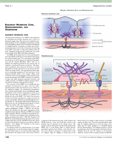
Plate 5-1                                                                                             Integumentary System

                                                                        BASEMENT MEMBRANE ZONE AND HEMIDESMOSOME
                                                  Basement membrane zone


                                                                       Keratinocyte
       BASEMENT MEMBRANE ZONE,                                                                               Hemidesmosome
       HEMIDESMOSOME, AND
       DESMOSOME

                                                                                                             Desmosome
       BASEMENT MEMBRANE ZONE
       The basement membrane zone (BMZ) of the epidermis
       is  a  beautiful  and  complex  structure  and  a  marvel  of                                        Lamina lucida
       biological engineering. The zone acts to attach the over-                                                         Basal lamina
       lying epidermis to the underlying stromal tissue, in this                                            Lamina densa
       case the papillary dermis, which is made predominantly
       of collagen bundles. A plethora of unique and special-                                               Reticular lamina
       ized proteins play critical roles in the proper functioning
       of the BMZ. Any defect or abnormal antibody that can
       cause disruption of the normal architecture can result
       in fracturing of the BMZ and blister formation.
         The BMZ can be appreciated on routine hematoxylin
       and eosin (H&E) staining as an eosinophilic band below   Hemidesmosome
       the basilar keratinocytes. The components of the BMZ
       are produced in two locations: the epidermal keratino-                  Hemidesmosome
       cyte  and  the  dermal  fibroblast.  These  cells  act  to
       produce  the  required  proteins  in  the  correct  ratio  to   Keratin
       maintain a functional basement membrane. The base-         5 and 14
       ment membrane’s most important function is to keep
       the epidermis firmly attached to the underlying dermis.   Cytoplasm
       This is necessary for life. This specialized structure also   of basal cell                                      Actin
       acts to encourage migration of cells and repair of the                                                 Kindlin
       epidermal-dermal  barrier  after  trauma.  Many  other
       critical processes and physiological roles depend on the           Plectin      BP230                           Actinin
       proper functioning of the BMZ, including permeability
       of  water  and  other  chemical  substrates,  proteins,  and                      BP180    CD151    Talin       Vinculin
       cellular elements. The BMZ is a highly organized struc-
       ture that is consistent from person to person.  Cell membrane
         The  structure  of  the  BMZ  can  be  subdivided  into   Lamina lucida   6  4                                  3  1
       individual  compartments  for  study,  with  the  under-        Integrin                                         Integrin
       standing that the entire unit functions as one. These are
       the  epidermal  basilar  cell  cytoskeleton,  hemidesmo-        Laminin 5                                     Laminin 10
       some, lamina lucida, lamina densa, and sublamina papil-  Lamina densa                   Collagen IV
       lary dermis. Each of these components is made up of
       unique  proteins  that  act  in  harmony  to  preserve  the   Nidogen
       functional role of the BMZ. The basilar keratinocytes
       contain intracellular cytoskeleton components made of                                Perlecan
       keratin intermediate filaments, predominantly keratin 5   Reticular lamina
       and keratin 14. The keratin intermediate filaments are                                     Anchoring fibrils
       interwoven into the hemidesmosomal plaque to firmly
       adhere the basilar cell to the hemidesmosome.
         The  keratin  intermediate  filaments  interact  with
       bullous  pemphigoid  antigen  1  (BP230)  and  plectin.         Collagen VII
       These  two  proteins  are  the  main  components  of  the
       hemidesmosomal plaque. Plectin and BP230 are bound
       tightly together. Plectin and bullous pemphigoid anti-
       body 1 also bind to the integrin class of proteins and to
       bullous pemphigoid antigen 2 (BP180). Integrins and
       BP180 are transcellular proteins that bind to the intra-
       cellular molecules, plectin and BP230; they also extend   composed of the transversing parts of the integrin and   lamina densa. It is unique in that it retains its globular
       out from the basilar keratinocyte and interact with the   BP180  proteins.  These  two  molecules  attach  to  the   regions on either end. These form attachments to other
       laminin  5  and  collagen  IV  molecules  in  the  lamina   laminin  class  of  proteins  in  the  lamina  densa.  The   type IV collagen molecules to create the lattice. Col-
       lucida and lamina densa.                  lamina lucida is considered to be the weakest part of the   lagen  type  IV  binds  strongly  to  a  dumbbell-shaped
         The lamina lucida is so named because of its trans-  BMZ,  and  it  is  the  blister  plane  in  suction  blisters,   protein named nidogen. This nidogen protein is critical
       lucent appearance on electron microscopy. In compari-  junctional  epidermolysis  bullosa,  and  salt-split  skin.   in attaching to the laminin proteins in the lamina densa.
       son, the lamina densa is an electron-dense region that   The lamina densa is composed of a latticework of type   Nidogen  locks  the  type  IV  collagen  to  the  laminins,
       lies just below the lamina lucida. The lamina lucida is   IV  collagen.  Type  IV  collagen  is  found  only  in  the   which are bound to the overlying integrin and BP180.

       150                                                                                   THE NETTER COLLECTION OF MEDICAL ILLUSTRATIONS