
Page 162 - The Netter Collection of Medical Illustrations - Integumentary System_ Volume 4 ( PDFDrive )
P. 162
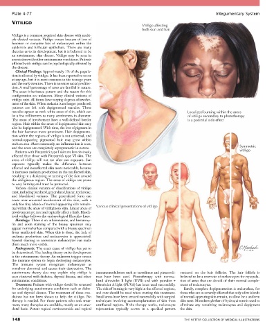
Plate 4-77                                                                                            Integumentary System
       VITILIGO
                                                                        Vitiligo affecting
                                                                        both skin and hair
       Vitiligo is a common acquired skin disease with multi-
       ple clinical variants. Vitiligo occurs because of loss of
       function  or  complete  loss  of  melanocytes  within  the
       epidermis  and  follicular  epithelium.  There  are  many
       theories as to its development, but it is believed to be
       an  autoimmune  skin  disease.  Vitiligo  may  be  seen  in
       association with other autoimmune conditions. Patients
       afflicted with vitiligo can be psychologically affected by
       the disease.
         Clinical Findings: Approximately 1% of the popula-
       tion is affected by vitiligo. It has been reported to occur
       at any age, but it is most common in the teenage years
       and the early twenties. There is no sex or racial predilec-
       tion. A small percentage of cases are familial in nature.
       The exact inheritance pattern and the reason for this
       configuration  are  unknown.  Many  clinical  variants  of
       vitiligo exist. All forms have varying degrees of involve-
       ment of the skin. When melanin is no longer produced,
       patients  are  left  with  depigmented  macules.  These
       macules appear as stark white areas of skin, which can                                  Localized burning within the areas
       be a few millimeters to many centimeters in diameter.                                   of vitiligo secondary to phototherapy
       The areas of involvement have a well-defined border                                     is a potential side effect
       region. Hair within the areas of depigmented skin may
       also be depigmented. With time, the loss of pigment in
       the hair becomes more prominent. Hair depigmenta-
       tion within the regions of vitiligo is not universal, and
       normal-appearing  pigmented  hair  may  grow  within
       such an area. Most commonly, no inflammation is seen,
       and the areas are completely asymptomatic in nature.                                                              Symmetric
         Patients with Fitzpatrick type I skin are less obviously                                                        vitiligo
       affected than those with Fitzpatrick type VI skin. The
       areas  of  vitiligo  will  not  tan  after  sun  exposure.  Sun
       exposure  typically  makes  the  difference  between
       affected and nonaffected skin more noticeable, because
       it increases melanin production in the unaffected skin,
       resulting in a darkening or tanning of the skin around
       the vitiliginous region. The areas of vitiligo are prone
       to easy burning and must be protected.
         Various clinical variants or classifications of vitiligo
       exist, including localized, generalized, linear, trichrome,
       and  blaschkoid  variants.  The  generalized  form  can
       cause  near-universal  involvement  of  the  skin,  with  a
       only few tiny islands of normal-appearing skin remain-  Various clinical presentations of vitiligo
       ing within the areas of vitiliginous skin. Linear areas of
       involvement are rare and typically affect a limb. Blasch-
       koid vitiligo follows the embryological Blaschko lines.
         Histology: There is no inflammation, and hematoxy-
       lin  and  eosin  staining  of  the  biopsy  specimen  may
       appear normal unless compared with a biopsy specimen
       from  unaffected  skin.  When  this  is  done,  the  lack  of
       melanin  production  and  melanocytes  is  appreciated.
       Special  staining  to  accentuate  melanocytes  can  make
       them much more visible.
         Pathogenesis: The exact cause of vitiligo has yet to
       be determined. The leading theory on its development
       is the autoimmune theory. An unknown trigger causes
       the immune system to begin destroying melanocytes.
       The  immune  system  recognizes  melanocytes  as
       somehow abnormal and causes their destruction. The
       autoimmune  theory  also  may  explain  why  vitiligo  is   immunomodulators such as tacrolimus and pimecroli-  centered  on  the  hair  follicles.  The  hair  follicle  is
       seen clustered with diabetes, thyroid disease, and other   mus  have  been  used.  Phototherapy  with  narrow-   believed to be a reservoir of melanocytes for repopula-
       autoimmune conditions.                    band  ultraviolet  B  light  (UVB)  and  with  psoralen  +   tion of areas that are devoid of their normal comple-
         Treatment: Patients with vitiligo should be screened   ultraviolet A light (PUVA) has been used successfully.   ment of melanocytes.
       for underlying autoimmune conditions such as diabe-  The risk of burning is very high in the affected regions,   Rarely, complete depigmentation is undertaken, for
       tes  and  thyroid  disease.  The  treatment  of  these  con-  and care should be used when starting this treatment.   those who are so severely affected that only a few islands
       ditions  has  not  been  shown  to  help  the  vitiligo.  No   Small areas have been treated successfully with surgical   of normal-appearing skin remain, to allow for a uniform
       therapy is needed. For those patients who seek treat-  techniques involving autotransplantation of skin from   skin tone. Monobenzylether of hydroquinone is used to
       ment, many therapies are available, mostly on an anec-  unaffected  regions.  If  the  therapy  works,  melanocyte   eliminate  any  remaining  melanocytes  and  depigment
       dotal  basis.  Potent  topical  corticosteroids  and  topical   rejuvenation  typically  occurs  in  a  speckled  pattern   the skin.

       148                                                                                   THE NETTER COLLECTION OF MEDICAL ILLUSTRATIONS