
Page 157 - The Netter Collection of Medical Illustrations - Integumentary System_ Volume 4 ( PDFDrive )
P. 157
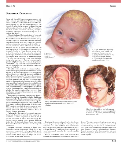
Plate 4-72                                                                                                           Rashes


        SEBORRHEIC DERMATITIS


        Seborrheic dermatitis is a commonly encountered rash
        with a bimodal age distribution. There is an infantile
        and an adult form. The two forms do not resemble each
        other  clinically  and  are  distinct  in  appearance.  The
        infantile form has also been named “cradle cap” because
        of its prominent location on the scalp. The adult form
        has  been  found  in  association  with  many  underlying
        conditions,  although  it  is  most  commonly  seen  as  an
        isolated skin finding.
          Clinical Findings: The infantile form of seborrheic
        dermatitis manifests in the first weeks of life and lasts a
        few months at most. It affects males and females equally,
        and there is no racial predilection. The most usual loca-
        tion of involvement is the scalp. Most cases are mild
        and  do  not  cause  the  parents  to  seek  the  advice  of  a
        medical professional. These mild cases manifest with a
        fine scale that may be slightly greasy or adherent. The
        child is unaware of the dermatosis, and it resolves spon-                                        In infants, seborrheic dermatitis
        taneously.  Rarely,  an  infant  develops  greasy  yellow,                                       is is referred to as “cradle cap”
        scaly patches and even plaques across the entire scalp                                           because of the development
        (cradle cap). The dermatitis may become more inflamed,                                           of greasy crusted patches on the
        and weeping from the patches or plaques may ensue.                                               scalp.This common finding
        The infant may try to scratch at the areas, indicating                                           in infancy typically improves
        that pruritus is present. In these severe cases, weeping                                         spontaneously over time.
        patches and plaques may also be seen in the groin and
        axillary folds. Only in the most exceptional of cases does
        the rash disseminate, but it has the ability to affect any
        region of the body.
          The adult version is chronic in nature and affects a
        higher percentage of people than does the infantile form.
        Because  of  its  chronicity,  patients  often  seek  medical
        advice. There is also quite a bit of clinical variability in
        adult seborrheic dermatitis. The face is the most com-
        monly involved site, with a predilection for the nasola-
        bial fold, eyebrows, ears, and scalp. It has a strikingly
        similarity to patches in other locations on the skin. Most
        cases are mild and consist of greasy yellow to slightly red,
        scaly patches. The scalp involvement is similar in appear-
        ance. Seborrheic dermatitis has a propensity to affect the
        areas of the skin that have a high density of sebaceous
        glands.  On  occasion,  patients  have  not  only  facial
        involvement but signs of involvement on areas of the
        upper chest and back.
          Many conditions have been associated with the adult
        form  of  seborrheic  dermatitis,  including  Parkinson’s
        disease and other chronic neurological disorders. Adult
        onset of severe seborrheic dermatitis has been reported   Severe seborrheic dermatitis may be associated
        to occur with a higher incidence in patients with under-  with human immunodeficiency virus (HIV)
        lying human immunodeficiency virus (HIV) infection.   infection.
        HIV-associated seborrheic dermatitis tends to be wide-
        spread,  with  severe  facial  involvement.  Patients  who
        present  with  severe  seborrheic  dermatitis  should  be                                 Seborrheic dermatitis in adults frequently
        assessed for HIV risk factors.                                                            manifests with greasy yellow, scaly patches
          Pathogenesis:  The  exact  pathogenesis  is  unknown.                                   in the scalp, ears, and eyebrows and along
        Seborrheic  dermatitis  is  believed  to  be  caused  by  an                              the nasolabial fold.
        interaction of various components of the skin, includ-
        ing  the  production  of  sebum,  with  the  normal  skin
        immune  system  response  to  the  fungus,  Malassezia
        furfur. The role that each of these factors plays in the   Treatment: Most cases of infantile seborrheic derma-  therapy. The other azole antifungal agents are just as
        formation  of  seborrheic  dermatitis  is  not  completely   titis can be ignored or treated with nothing more than   effective. The addition of a weak topical corticosteroid
        understood.                               daily baths and a bland emollient. More involved cases   used  intermittently  can  also  lead  to  excellent  results.
          Histology:  Seborrheic  dermatitis  is  almost  never   can be treated with more frequent shampooing of the   The scalp is most commonly treated with a ketoconazole-
        biopsied to confirm the diagnosis. Classic biopsy spe-  scalp and the use of a mild topical corticosteroid. The   based shampoo or a tar- or selenium-based shampoo.
        cimens show parakeratosis overlying a slightly spongi-  use of ketoconazole cream has also been advocated in   There  is  no  cure  for  seborrheic  dermatitis,  but  most
        otic  epidermis  with  a  mild  lymphocytic  perivascular   some cases.             therapeutic regimens, if adhered to, lead to an excellent
        infiltrate in the dermis. Spores of fungus can be seen   Because of its chronic nature, adult seborrheic der-  clinical response.
        lying on the surface of the epidermis.    matitis is treated with topical ketoconazole as a first-line


        THE NETTER COLLECTION OF MEDICAL ILLUSTRATIONS                                                                          143