
Page 154 - The Netter Collection of Medical Illustrations - Integumentary System_ Volume 4 ( PDFDrive )
P. 154
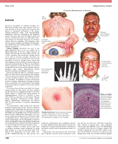
Plate 4-69                                                                                            Integumentary System

                                                                           CUTANEOUS MANIFESTATIONS OF SARCOID


                                                  Skin
                                                  lesions
       SARCOID


       Sarcoid,  or  sarcoidosis,  is  a  relatively  common  con-
       dition that can affect many organ systems. There is a
       wide  spectrum  of  disease  activity,  from  localized  skin
       disease  to  widespread  involvement  of  the  integu-
       mentary,  pulmonary,  cardiac,  renal,  gastrointestinal,                                                     Bilateral
       ophthalmic,  endocrine,  neurological,  and  lymphatic                                                        parotid gland
       systems. However, most cases are mild in nature and                                                           involvement
       can be controlled with proper care. Although an infec-
       tious etiology has often been theorized, no conclusive   Lacrimal
       evidence has been established. This idiopathic condi-  gland involve-
       tion can produce multiple skin findings. The skin find-  ment
       ings should cause the attending physician to look for
       systemic involvement.
         Clinical  Findings:  Sarcoidosis  can  occur  in  any
       ethnic  population,  but  it  is  seen  at  a  higher  rate  in
       African  Americans.  It  also  has  a  higher  incidence  in
       women. The usual age at onset is before 40 years. Up
       to 90% of patients with sarcoid have a benign clinical
       course  with  no  increased  mortality.  Sarcoidosis  has
       been reported to occur in a familial form, which has led
       researchers  to  look  for  specific  genetic  defects  that                                                    Paralysis due
       could explain the disease. However, sarcoid remains an                                                          to involvement
       idiopathic multisystem disease. There are many distinct                                                         of facial
       clinical  expressions  of  the  disease  that  are  common                                                      (VII) nerve
       enough that they have been named, including Löfgren’s
       syndrome,  lupus  pernio,  Darier-Roussy  sarcoid,     Bone destruction
       Heerfordt syndrome, and Mikulicz syndrome.  of terminal
         Sarcoid  can  affect  the  skin  in  a  multitude  of  ways.   phalanges
       There are both specific and nonspecific skin findings.
       The most common nonspecific skin finding is erythema
       nodosum. Erythema nodosum affects the lower ante-
       rior  extremities.  It  manifests  as  tender  subcutaneous
       nodules or plaques. Examination of biopsy specimens
       shows a nonspecific form of panniculitis. The etiology
       of erythema nodosum in patients with sarcoid is poorly
       understood.
         The lesions of sarcoid that occur within the integu-
       mentary  system  are  quite  varied.  The  most  common
       specific skin lesion is a slightly brownish to red-brown
       papule,  plaque,  or  nodule  with  varying  amounts  of                                                      Biopsy of nodule.
       hyperpigmentation.  Sarcoid  is  a  mimicker  of  many                                                        Typical sarcoidal
       other conditions, especially in its skin lesions. Macular                                                     granuloma (dense
       lesions,  ulcerations,  subcutaneous  nodules,  annular                                                       infiltration with
       plaques,  ichthyosiform  erythroderma,  and  alopecia                                                         macrophages,
       have  all  been  described  as  potential  presentations                                                      epithelioid cells,
       of sarcoid.                                                                                                   and occasional
         The  extracutaneous  organ  system  most  commonly                                                          multinucleated
       involved is the pulmonary system. There is a relatively                                                       giant cells [arrow])
       straightforward classification that describes the stages
       of pulmonary sarcoid based on radiographic findings.
       The higher the radiographic stage, the more severe the   Positive Kveim test. Intracutaneous injection
       disease. Isolated bilateral hilar adenopathy is the most   of saline suspension of human sarcoidal
       common pulmonary finding, and it is the basis for stage   spleen or lymph nodes causes appearance
       I  radiographic  disease.  These  patients  are  most  com-  of erythematous nodule in 2 to 6 weeks.
       monly asymptomatic, and the adenopathy is found on
       routine radiographic testing. Any findings of pulmonary
       sarcoid should prompt referral of the affected individual   erythrocyte  sedimentation  rate  is  uniformly  elevated.   nose and the rest of the face. This form of sarcoid is
       to a pulmonologist for pulmonary function testing.  For some unknown reason, this syndrome is most com-  quite  resistant  to  therapy,  runs  a  more  prolonged
         Löfgren’s  syndrome  is  defined  by  the  acute  onset     monly seen in young Caucasian women. This form of   course, and is often difficult to treat. The skin findings
       of  erythema  nodosum,  almost  exclusively  in  young   sarcoidosis typically resolves spontaneously within 2 to   are  typically  shiny  brown-red  plaques,  papules,  and
       adult  women;  it  is  seen  in  association  with  fever,     3 years.             nodules  overlying  the  nose  and  other  regions  of  the
       bilateral  hilar  adenopathy,  and  uveitis.  Other,  non-  Lupus pernio is the name given to the clinical find-  face. The involvement can become so severe as to cause
       specific  constitutional  signs  are  often  present.  The   ings  of  specific  cutaneous  sarcoid  involvement  of  the   disfigurement of the nose by shiny red-brown papules

       140                                                                                   THE NETTER COLLECTION OF MEDICAL ILLUSTRATIONS