
Page 193 - The Netter Collection of Medical Illustrations - Integumentary System_ Volume 4 ( PDFDrive )
P. 193
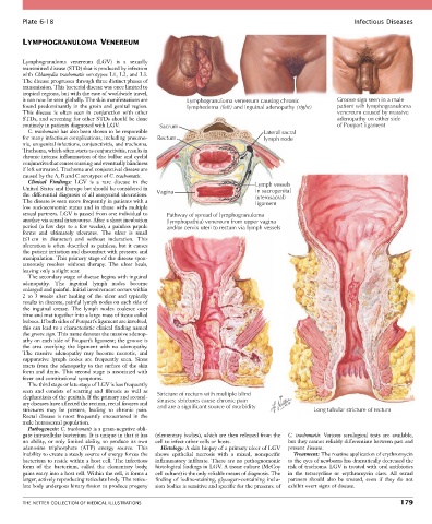
Plate 6-18                                                                                               Infectious Diseases


        LYMPHOGRANULOMA VENEREUM


        Lymphogranuloma  venereum  (LGV)  is  a  sexually
        transmitted disease (STD) that is produced by infection
        with Chlamydia trachomatis serotypes L1, L2, and L3.
        The disease progresses through three distinct phases of
        transmission. This bacterial disease was once limited to
        tropical regions, but with the ease of worldwide travel,
        it can now be seen globally. The skin manifestations are   Lymphogranuloma venereum causing chronic  Groove sign seen in a male
        found predominantly in the groin and genital region.   lymphedema (left) and inguinal adenopathy (right)  patient wih lymphogranuloma
        This  disease  is  often  seen  in  conjunction  with  other                                        venereum caused by massive
        STDs, and screening for other STDs should be done                                                   adenopathy on either side
        routinely in patients diagnosed with LGV.   Sacrum                                                  of Poupart ligament
          C. trachomatis has also been shown to be responsible                      Lateral sacral
        for many infectious complications, including pneumo-  Rectum                lymph node
        nia, urogenital infections, conjunctivitis, and trachoma.
        Trachoma, which often starts as conjunctivitis, results in
        chronic intense inflammation of the bulbar and eyelid
        conjunctiva that causes scarring and eventually blindness
        if left untreated. Trachoma and conjunctival disease are
        caused by the A, B and C serotypes of C. trachomatis.
          Clinical  Findings:  LGV  is  a  rare  disease  in  the                 Lymph vessels
        United States and Europe but should be considered in                      in sacrogenital
        the differential diagnosis of all anogenital ulcerations.   Vagina        (uterosacral)
        The disease is seen more frequently in patients with a                    ligament
        low  socioeconomic  status  and  in  those  with  multiple
        sexual partners. LGV is passed from one individual to   Pathway of spread of lymphogranuloma
        another via sexual intercourse. After a short incubation   (lymphopathia) venereum from upper vagina
        period (a few days to a few weeks), a painless papule   and/or cervix uteri to rectum via lymph vessels
        forms  and  ultimately  ulcerates.  The  ulcer  is  small
        (≤1 cm  in  diameter)  and  without  induration.  This
        ulceration is often described as painless, but it causes
        the patient irritation and discomfort with pressure and
        manipulation. This primary stage of the disease spon-
        taneously  resolves  without  therapy.  The  ulcer  heals,
        leaving only a slight scar.
          The secondary stage of disease begins with inguinal
        adenopathy.  The  inguinal  lymph  nodes  become
        enlarged and painful. Initial involvement occurs within
        2  to  3  weeks  after  healing  of  the  ulcer  and  typically
        results in discrete, painful lymph nodes on each side of
        the  inguinal  crease.  The  lymph  nodes  coalesce  over
        time and mat together into a large mass of tissue called
        buboes. If both sides of Poupart’s ligament are involved,
        this can lead to a characteristic clinical finding named
        the groove sign. This name denotes the massive adenop-
        athy on each side of Poupart’s ligament; the groove is
        the  area  overlying  the  ligament  with  no  adenopathy.
        The  massive  adenopathy  may  become  necrotic,  and
        suppurative  lymph  nodes  are  frequently  seen.  Sinus
        tracts from the adenopathy to the surface of the skin
        form  and  drain.  This  second  stage  is  associated  with
        fever and constitutional symptoms.
          The third stage or late stage of LGV is less frequently
        seen  and  consists  of  scarring  and  fibrosis  as  well  as   Stricture of rectum with multiple blind
        elephantiasis of the genitals. If the primary and second-  sinuses; strictures cause chronic pain
        ary diseases have affected the rectum, rectal fissures and   and are a significant source of morbidity
        strictures  may  be  present,  leading  to  chronic  pain.                                  Long tubular stricture of rectum
        Rectal  disease  is  most  frequently  encountered  in  the
        male homosexual population.
          Pathogenesis: C. trachomatis is a gram-negative obli-
        gate intracellular bacterium. It is unique in that it has   (elementary bodies), which are then released from the   C.  trachomatis.  Various  serological  tests  are  available,
        no  ability,  or  only  limited  ability,  to  produce  its  own   cell to infect other cells or hosts.  but they cannot reliably differentiate between past and
        adenosine  triphosphate  (ATP)  energy  source.  This   Histology: A skin biopsy of a primary ulcer of LGV   present disease.
        inability to create a steady source of energy forces the   shows  epithelial  necrosis  with  a  mixed,  nonspecific   Treatment: The routine application of erythromycin
        bacterium to reside within a host cell. The infectious   inflammatory infiltrate. There are no pathognomonic   to the eyes of newborns has dramatically decreased the
        form  of  the  bacterium,  called  the  elementary  body,   histological findings in LGV. A tissue culture (McCoy   risk of trachoma. LGV is treated with oral antibiotics
        gains entry into a host cell. Within the cell, it forms a   cell culture) is the only reliable means of diagnosis. The   in  the  tetracycline  or  erythromycin  class.  All  sexual
        larger, actively reproducing reticulate body. The reticu-  finding of iodine-staining, glycogen-containing inclu-  partners  should  also  be  treated,  even  if  they  do  not
        late body undergoes binary fission to produce progeny   sion bodies is sensitive and specific for the presence of   exhibit overt signs of disease.


        THE NETTER COLLECTION OF MEDICAL ILLUSTRATIONS                                                                          179