
Page 188 - The Netter Collection of Medical Illustrations - Integumentary System_ Volume 4 ( PDFDrive )
P. 188
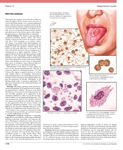
Plate 6-13                                                                                            Integumentary System


                                                        Ulcerating plaque of tongue
       HISTOPLASMOSIS                                   due to histoplasmosis. Lesion
                                                        may be identical in appearance
                                                        to carcinoma of tongue.
       Histoplasmosis is endemic in the Ohio River Valley but
       exists  throughout  North  America  and  is  also  seen  in
       Central and South America. It is a primary pulmonary
       disease, with the skin being secondarily involved in dis-
       seminated disease; however, isolated cutaneous disease
       can result from direct inoculation. The disease is typi-
       cally  seen  in  immunocompromised  patients.  Patients
       typically breathe in the infective spores, which lodge in
       the pulmonary tree. Most infections are subclinical.
         Clinical  Findings: The disease is seen primarily in
       immunocompromised  patients.  Other  risk  factors
       include occupations that increase the patient’s contact
       with  bat  or  bird  droppings  in  an  endemic  region.
       The  fungus  is  not  found  within  bird  droppings,  but
       the droppings provide the perfect environment for the
       fungus  to  grow  and  reproduce.  Patients  inhale  the
       spores into the lungs. Most have no symptoms. Some
       have  mild  flu-like  symptoms  that  go  undiagnosed  or
       misdiagnosed  as  an  upper  respiratory  infection.  The
       primary infection heals, and the lungs may have visible
       findings  on  chest  radiography.  Variable  radiographic
       findings are seen. Small, symmetrically located areas of
       hilar miliary calcification are the most common finding.
       Other  lung  findings  can  mimic  those  of  tuberculosis,
       lung  cancer,  or  metastatic  cancer.  Bilateral  hilar  ade-
       nopathy may be seen, as may lobar pneumonia.
         Dissemination  of  the  disease  to  other  organs  can   Mycelial or free-living phase of H. capsulatum as it
       occur  in  the  immunocompromised  host.  The  skin  is   exists in nature or in culture
       commonly  affected  in  disseminated  disease.  The  skin
       findings  often  appear  as  papules,  plaques,  or  nodules
       with  varying  degrees  of  ulceration.  Subcutaneous
       abscess formation may occur, and fistulas and sinus tract                                    Spores of mycelial phase of
       formation may be prominent. Surrounding redness may                                          H. capsulatum. Inhalation of these
       give  the  appearance  of  cellulitis.  Adenopathy  in  the                                  is the source of infection.
       draining  lymph  nodes  is  commonly  appreciated.  The
       diagnosis is dependent on the histological findings and
       the culture results.
         Histology: Skin biopsy specimens show pseudocarci-
       nomatous hyperplasia of the epidermis with an underly-
       ing  granulomatous  infiltrate.  Ulceration  and  abscess
       formation are not uncommon with widespread necrosis.
       The  yeast-like  organisms  can  be  appreciated  in  the
       cytoplasm of histiocytes. This is one of the few infec-
       tions in which one sees phagocytized histiocytes. The
       yeast structures are round to oval, and there may be a
       clear region surrounding the yeast cell. Yeast organisms
       are  also  appreciated  within  the  dermis,  between  and
       within the inflammatory infiltrate. They can be high-
       lighted  by  use  of  special  histology  stains  such  as  the
       periodic acid–Schiff stain or the Grocott silver stain.
         The fungus is best cultured on Sabouraud’s media.
       The fungus in its mycelial phase grows slowly. It appears   Dimorphic fungus. H. capsulatum in tissue  H. capsulatum in a macrophage, termed
       as a brown, fluffy fungus on culture.                                                     a phagocytized histiocyte. In this yeast or
         Pathogenesis: Histoplasma capsulatum is a dimorphic                                     tissue phase, the organism is not
       fungus that is responsible for a wide range of infections                                 transmissible from person to person.
       including  pulmonary,  pericardial,  and  cutaneous  dis-
       eases. The fungus is ubiquitous in nature and is found
       in soil, where it lives as a saprophyte. Spores from the
       mycelial phase of the fungus are inhaled or inoculated   preexposed or newly exposed patient becomes immu-  immunocompromised  should  be  started  on  therapy
       directly into the skin. Once they have entered the body,   nosuppressed, the patient is at risk for disease reactiva-  with one of the three most efficacious and best-studied
       the change in temperature causes transformation of the   tion and serious sequelae.  medications: fluconazole, itraconazole, or amphotericin
       spores into the yeast form of H. capsulatum. Most infec-  Treatment: Most cases of primary pulmonary disease   B.  Treatment  may  be  prolonged.  Patients  who  are
       tions  go  unnoticed,  and  most  of  the  others  induce  a   go undiagnosed, and the patient’s immune system con-  found  to  have  the  acquired  immunodeficiency  syn-
       subclinical  scenario  or  a  mild,  flu-like  illness.  Most   tains the fungus. In those patients with mild pulmonary   drome benefit from directed therapy against the human
       cases  are  self-contained,  and  the  only  evidence  of   symptoms who are not immunocompromised, therapies   immunodeficiency virus. Patients taking chronic immu-
       disease is the formation of granulomas within the lungs   can  be  withheld,  because  most  cases  resolve  spon-  nosuppressants should have their medications discon-
       and  a  positive  skin  delayed-hypersensitivity  test.  If  a   taneously. Patients who have more severe disease or are   tinued or decreased, if possible.

       174                                                                                   THE NETTER COLLECTION OF MEDICAL ILLUSTRATIONS