
Page 196 - The Netter Collection of Medical Illustrations - Integumentary System_ Volume 4 ( PDFDrive )
P. 196
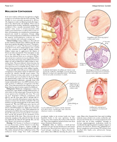
Plate 6-21                                                                                            Integumentary System


       MOLLUSCUM CONTAGIOSUM


       As its name implies, molluscum contagiosum is a highly
       contagious viral infection that has little morbidity. This
       infection is most commonly encountered in children.
       The diagnosis is made on clinical grounds after inspec-
       tion of the characteristic skin findings. When seen in
       the genital region of adults, molluscum contagiosum is
       considered  to  be  a  sexually  transmitted  disease.  This
       infection  rarely  occurs  in  immunocompetent  adults
       outside sexual transmission. In adults with no clear evi-
       dence of transmission, an evaluation for an immunosup-
       pressed  state  should  be  undertaken.  Patients  taking
       chronic immunosuppressive medications and those with
       the  acquired  immunodeficiency  syndrome  are  more                                            Magnified view showing typical
       prone to infection with molluscum contagiosum.                                                  umbilicated lesion
         Clinical Findings: Young children are often affected
       by this common viral infection. Children pass the virus                                                              Keratin
                                                                                                                            plug
       from one to another through close contact. The incuba-
       tion period is 2 to 4 weeks. The characteristic finding is
       of small (3-5 mm), dome-shaped papules with a central
       dell.  The  coloration  can  be  pink  to  slightly  whitish.
       Solitary  lesions  may  be  appreciated,  but  clusters  of
       lesions are often encountered. They may appear on any
       part  of  the  body.  Slight  pruritus  may  accompany  the
       lesions, but otherwise there are no symptoms. Mollus-
       cum lesions have a tendency to become inflamed. When
       this occurs, they can become tender. Inflamed lesions are
       bright red and can bleed if the child scratches or trau-
       matizes them. The more inflamed a lesion becomes, the                                  Inclusion
       more likely it is to leave scarring. Scarring can also occur                           bodies
       if the lesion becomes secondarily infected. Most nonin-
       flamed lesions spontaneously resolve within 6 months.
         Young and older adults who present with molluscum   Scattered distribution of molluscum lesions over  Histological section of molluscum
       contagiosum in the genital region are believed to have   perineum, buttocks, and thighs. Lesions spread by  lesions showing poxvirus inclusion
       acquired  the  infection  through  sexual  contact.  The   physical contact and autoinoculation. The disease  bodies and central core of keratin
       number of lesions in these cases tends to be increased,   can be sexually transmitted in adults.
       and the lesions tend to be localized to the groin. These
       also spontaneously resolve over time with no therapy.
       Immunosuppressed  individuals,  especially  those  with
       human immunodeficiency virus (HIV) infection, have a
       high incidence of molluscum contagiosum viral infec-                       Application of
       tions. These infections tend to be widespread and can be                   liquid nitrogen
       larger than the typical version acquired in childhood.                     to lesion using
         Pathogenesis: Molluscum contagiosum is caused by                         cotton swab
       an enveloped, large, double-stranded DNA poxvirus, of
       which there are four unique types. Humans are the only
       known species to be infected by this virus. The virus
       has  been  designated  molluscum  contagiosum  virus
       (MCV),  and  the  four  types  MCV1  through  MCV4.
       The virus is spread by close physical contact, and trans-
       mission on fomites has also been established. The virus
       attaches to the glycosaminoglycans on the surface of the
       targeted cell. The viral DNA gains entry into the cell                   with
       cytoplasm, where it replicates itself. The virus carries                 E. Hatton
       with it a viral RNA polymerase, which acts to transcribe   Local eradication of lesions can be obtained  Molluscum contagiosum
       the viral genes, as well as a viral DNA polymerase for   with desiccation, cryotherapy, laser ablation,  is commonly encountered
       replication of its DNA. Early and late proteins are pro-  chemical cautery, or curettage.          in children.
       duced. The early proteins are generally for viral replica-
       tion,  and  the  late  proteins  are  for  production  of  the
       structural shell of the virus. These processes all occur   cytoplasmic  bodies  in  the  stratum  basalis  into  larger   cause. Many other therapies have been used, including
       within the cytoplasm of the infected cell. Once the virus   basophilic  bodies  in  the  outer  epidermis.  As  they   tretinoin cream, salicylic acid, curette, cantharidin, and
       has replicated, the infected cell typically dies, and the   enlarge, they often compress the nucleus of the infected   imiquimod. Patients who are immunosuppressed can be
       brick-shaped viral particles are released.  cell. These intracytoplasmic inclusion bodies have been   treated  with  any  of  these  modalities.  Attempts  to
         Histology: Skin biopsies of molluscum contagiosum   termed Henderson-Patterson bodies.  decrease  immunosuppressive  medications  should  be
       are very characteristic, and the infection is easily diag-  Treatment:  Often  in  children,  a  watch-and-wait   coordinated through the patient’s transplant surgeon or
       nosed  histologically.  However,  biopsies  usually  are     approach is the best therapy, because most cases resolve   primary care physician. Patients with widespread mol-
       not obtained because the disease is diagnosed clinically.   spontaneously. Many destructive methods are available.   luscum contagiosum and coexisting HIV infection have
       The virally infected cells have molluscum bodies. The   Liquid  nitrogen  cryotherapy  is  highly  effective,  but   benefited  from  highly  active  antiretroviral  therapy
       molluscum  bodies  change  from  small,  eosinophilic   most children have difficulty tolerating the pain it can   (HAART).

       182                                                                                   THE NETTER COLLECTION OF MEDICAL ILLUSTRATIONS