
Page 195 - The Netter Collection of Medical Illustrations - Integumentary System_ Volume 4 ( PDFDrive )
P. 195
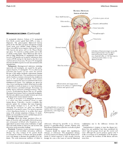
Plate 6-20                                                                                               Infectious Diseases

                                                                                   BACTERIAL MENINGITIS
                                                                                   Sources of infection
                                                               Basal skull fracture
                                                                                                         Cribriform plate defect
                                                                    Otitis media
                                                                                                         Sinusitis (ethmoiditis)
                                                                     Mastoiditis
                                                                                                         Nasal furuncles

        MENINGOCOCCEMIA (Continued)                                                                      Nasopharyngitis


        N.  meningitidis  infection.  Culture  of  N.  meningitidis                                      Pneumonia
        from  blood,  cerebral  spinal  fluid  (CSF),  or  tissue  is
        diagnostic.  The  gram-negative  diplococcal  bacteria
        grows on the chocolate agar plate and appears as small,
        round,  moist,  gray  colonies.  Gram  staining  of  CSF
        shows intracellular gram-negative diplococcal bacteria.   Dermal sinuses
        This  bacteria  also  grows  well  on  the  Thayer-Martin
        agar plate. The bacteria is oxidase positive and is able                                       Infection of leptomeninges is usually
        to acidify certain sugars. These laboratory data can be                                        hematogenous but may be direct
                                                                                                       from paranasal sinuses, middle
        used  to  help  differentiate  N.  meningitidis  from  other                                   ear, mastoid cells, or CSF leak due to
        bacteria. CSF samples can be used for polymerase chain                                         cribriform plate defect or via
        reaction (PCR) testing for the bacteria, but this is not                                       dermal sinuses.
        routinely done in these cases. All cases of N. meningitidis
        infection should be reported to state and national health
        organizations.
          Pathogenesis:  Meningococcal  infections,  including   Skin (furuncles)
        septicemia  and  meningitis,  are  caused  by  the  gram-
        negative bacteria, N. meningitidis. This is a diplococcal
        bacterium  that  requires  an  iron  source  for  survival.
        Because of this unique metabolic requirement, humans
        are the only known host. The meningococcus bacteria
        can be found as a transient colonizer in the oropharynx
        of  up  to  10%  of  sampled  individuals.  These  carriers
        express no sequelae but serve as a potential reservoir for
        meningococcal  disease.  The  organisms  are  spread  by
        close contact and sharing of saliva. If the bacteria is able   Inflammation and suppurative
        to reproduce to such an extent as to cause bacteremia,   process on surface of leptomeninges
        it then becomes a potential pathogen. Bacteremia can    of brain and spinal cord
        quickly lead to septicemia (meningococcemia). This is
        a severe, life-threatening disease that can kill quickly.
        Meningeal involvement leads to neisserial meningitis.
        The bacteria exhibit a neurotrophic behavior and attack
        the lining of the central nervous system.
          At  least  13  serotypes  of  N.  meningitidis  are  known,
        9  of  which  have  been  conclusively  shown  to  cause
        human  disease.  Currently,  a  vaccine  is  available  that
        protects  against  the  serotypes  that  most  frequently
        cause  disease:  serotypes  A,  C,  Y,  and  W-135.  The
        remaining  five  serotypes  can  affect  any  individual   Thrombophlebitis of superior
        regardless of vaccination status. The bacteria expresses   sagittal sinus and suppurative
        a toxin (lipooligosaccharide) on its surface that causes   ependymitis, with beginning
        many of the systemic symptoms of disease. N. menin-  hydrocephalus
        gitidis is an encapsulated bacteria, and this helps protect
        it from the host’s immune system.
          Histology:  Most  skin  biopsy  specimens  show  evi-
        dence of vasculitis with neutrophils, fibrinoid necrosis,
        and  extravasated  red  blood  cells.  Organisms  can  be
        appreciated on tissue Gram stains. Embolism of capil-  ceftriaxone,  followed  by  penicillin  or  by  chloram-  confirmation  can  be  the  difference  between  life
        laries and small venules is often seen, and necrosis and   phenicol  in  penicillin-allergic  patients.  Patients  with   and death.
        ulceration can be secondary findings.     Waterhouse-Friderichsen syndrome need adrenal gland   Immunization  is  helping  to  keep  the  disease  inci-
          Treatment: Treatment requires prompt recognition   replacement therapy.           dence  low,  and  guidelines  have  been  established  for
        of  symptoms  and  immediate  intravenous  antibiotic   Contacts  should  be  treated  with  ciprofloxacin,   which  high-risk  groups  should  get  the  vaccine  and
        therapy.  Any  close  contacts  of  the  patient  should     rifampin,  or  ceftriaxone.  This  prophylactic  therapy,     when.  Although  the  vaccine  protects  against  only  4
        be screened for evidence of disease and given prophy-  as well as intravenous therapy, should be started imme-  of the 13 serotypes of N. meningitidis, it has the poten-
        lactic oral therapy to decrease the potential of an epi-  diately  if  clinical  suspicion  is  high  enough;  delaying   tial to decrease the incidence of this disease and save
        demic.  The  main  intravenous  antibiotic  of  choice  is   therapy  for  even  a  few  hours  to  wait  for  laboratory   many lives.


        THE NETTER COLLECTION OF MEDICAL ILLUSTRATIONS                                                                          181