
Page 197 - The Netter Collection of Medical Illustrations - Integumentary System_ Volume 4 ( PDFDrive )
P. 197
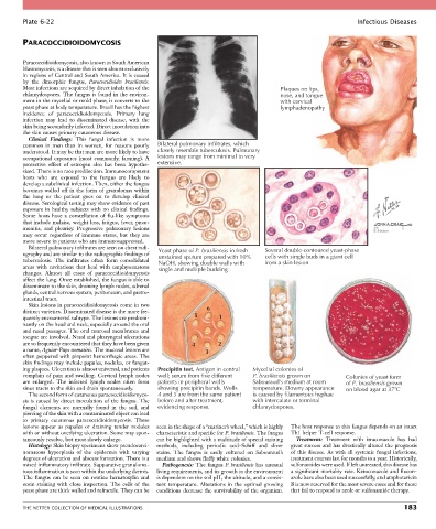
Plate 6-22                                                                                               Infectious Diseases


        PARACOCCIDIOIDOMYCOSIS


        Paracoccidioidomycosis, also known as South American
        blastomycosis, is a disease that is seen almost exclusively
        in regions of Central and South America. It is caused
        by  the  dimorphic  fungus,  Paracoccidioides  brasiliensis.
        Most infections are acquired by direct inhalation of the                         Plaques on lips,
        chlamydospores. The fungus is found in the environ-                              nose, and tongue
        ment in the mycelial or mold phase; it converts to the                           with cervical
        yeast phase at body temperature. Brazil has the highest                          lymphadenopathy
        incidence  of  paracoccidioidomycosis.  Primary  lung
        infection  may  lead  to  disseminated  disease,  with  the
        skin being secondarily infected. Direct inoculation into
        the skin causes primary cutaneous disease.
          Clinical  Findings:  This  fungal  infection  is  more
        common  in  men  than  in  women,  for  reasons  poorly   Bilateral pulmonary infiltrates, which
        understood. It may be that men are more likely to have   closely resemble tuberculosis. Pulmonary
        occupational  exposures  (most  commonly,  farming).  A   lesions may range from minimal to very
        protective  effect  of  estrogen  also  has  been  hypothe-  extensive.
        sized. There is no race predilection. Immunocompetent
        hosts  who  are  exposed  to  the  fungus  are  likely  to
        develop a subclinical infection. Then, either the fungus
        becomes walled off in the form of granulomas within
        the  lung  or  the  patient  goes  on  to  develop  clinical
        disease. Serological testing may show evidence of past
        exposure in healthy subjects with no clinical findings.
        Some hosts have a constellation of flu-like symptoms
        that include malaise, weight loss, fatigue, fever, pneu-
        monitis,  and  pleurisy.  Progressive  pulmonary  lesions                                                     with
                                                                                                                      E. Hatton
        may  occur  regardless  of  immune  status,  but  they  are
        more severe in patients who are immunosuppressed.
          Bilateral pulmonary infiltrates are seen on chest radi-                   Several double-contoured yeast-phase
        ography and are similar to the radiographic findings of   Yeast phase of P. brasiliensis in fresh  cells with single buds in a giant cell
                                                   unstained sputum prepared with 10%
        tuberculosis.  The  infiltrates  often  form  consolidated   NaOH, showing double walls with  from a skin lesion
        areas  with  cavitations  that  heal  with  emphysematous   single and multiple budding
        changes.  Almost  all  cases  of  paracoccidioidomycosis
        affect the lung. Once established, the fungus is able to
        disseminate to the skin, draining lymph nodes, adrenal
        glands, central nervous system, peritoneum, and gastro-
        intestinal tract.
          Skin lesions in paracoccidioidomycosis come in two
        distinct varieties. Disseminated disease is the more fre-  6
        quently encountered subtype. The lesions are predomi-  5      1
        nantly on the head and neck, especially around the oral
        and nasal passages. The oral mucosal membranes and
        tongue are involved. Nasal and pharyngeal ulcerations
        are so frequently encountered that they have been given   4  2
        a name, Aguiar-Pupo stomatitis. The mucosal lesions are   3
        often peppered with pinpoint hemorrhagic areas. The
        skin findings may include papules, nodules, or fungat-
        ing plaques. Ulceration is almost universal, and patients   Precipitin test. Antigen in central  Mycelial colonies of
        complain of pain and swelling. Cervical lymph nodes   well; serum from five different  P. brasiliensis grown on  Colonies of yeast form
        are  enlarged.  The  infected  lymph  nodes  often  form   patients in peripheral wells  Sabouraud’s medium at room  of P. brasiliensis grown
        sinus tracts to the skin and drain spontaneously.  showing precipitin bands. Wells  temperature. Downy appearance  on blood agar at 37 C
          The second form of cutaneous paracoccidioidomyco-  4 and 5 are from the same patient  is caused by filamentous hyphae
        sis is caused by direct inoculation of the fungus. The   before and after treatment,  with intercalate or terminal
        fungal  elements  are  normally  found  in  the  soil,  and   evidencing response.  chlamydospores.
        piercing of the skin with a contaminated object can lead
        to  primary  cutaneous  paracoccidioidomycosis.  These
        lesions  appear  as  papules  or  draining  tender  nodules   seen in the shape of a “mariner’s wheel,” which is highly   The host response to this fungus depends on an intact
        with or without overlying ulceration. Some may spon-  characteristic and specific for P. brasiliensis. The fungus   Th1 helper T-cell response.
        taneously resolve, but most slowly enlarge.  can be highlighted with a multitude of special staining   Treatment:  Treatment  with  itraconazole  has  had
          Histology: Skin biopsy specimens show pseudocarci-  methods,  including  periodic  acid–Schiff  and  sliver   great success and has drastically altered the prognosis
        nomatous  hyperplasia  of  the  epidermis  with  varying   stains.  The  fungus  is  easily  cultured  on  Sabouraud’s   of this disease. As with all systemic fungal infections,
        degrees of ulceration and abscess formation. There is a   medium and shows fluffy white colonies.  treatment courses last for months to a year. Historically,
        mixed inflammatory infiltrate. Suppurative granuloma-  Pathogenesis: The fungus P. brasiliensis has unusual   sulfonamides were used. If left untreated, this disease has
        tous inflammation is seen within the underlying dermis.   living requirements, and its growth in the environment   a significant mortality rate. Ketoconazole and flucon-
        The  fungus  can  be  seen  on  routine  hematoxylin  and   is dependent on the soil pH, the altitude, and a consis-  azole have also been used successfully, and amphotericin
        eosin  staining  with  close  inspection.  The  cells  of  the   tent  temperature.  Alterations  in  the  optimal  growing   B is now reserved for the most severe cases and for those
        yeast phase are thick walled and refractile. They can be   conditions decrease the survivability of the organism.   that fail to respond to azole or sulfonamide therapy.


        THE NETTER COLLECTION OF MEDICAL ILLUSTRATIONS                                                                          183