
Page 189 - The Netter Collection of Medical Illustrations - Integumentary System_ Volume 4 ( PDFDrive )
P. 189
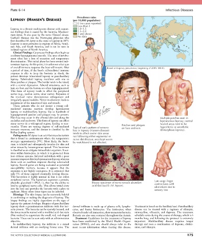
Plate 6-14                                                                                               Infectious Diseases

                                                     Prevalence rates
        LEPROSY (HANSEN’S DISEASE)                (per 10,000 population)
                                                      0 (no cases reported)
                                                      Less than 1
        Leprosy is a chronic multisystem disease with cutane-  1.0–1.5
        ous findings that is caused by the bacteria, Mycobacte-  1.5–2.0
        rium leprae. It also goes by the name Hansen’s disease.   2 and above
        Gerhard  Hansen  was  the  Norwegian  physician  who   No data
        first described M. leprae as the cause of leprosy in 1873.
        Leprosy is most prevalent in regions of Africa, South-
        east  Asia,  and  South  America,  and  it  can  be  seen  in
        isolated regions of North America.
          Clinical Findings: Cutaneous findings often begin as
        a solitary hypopigmented macule. The area of involve-
        ment  often  has  a  loss  of  sensation  and  temperature
        discrimination. This initial phase has been termed inde-
        terminate leprosy. At this point, it is unknown what type
        of overall immune response the host will mount. After            Based on Leprosy prevalence, beginning of 2009. WHO.
        a  period  of  time,  if  the  host’s  cell-mediated  immune
        response  is  able  to  keep  the  bacteria  in  check,  the
        patient develops tuberculoid leprosy or paucibacillary
        leprosy.  Tuberculoid  leprosy  manifests  with  one  to
        three patches or plaques. The border tends to be raised,
        with a central depression. Adnexal structures, such as
        hair, are lost, and the lesions are often hypopigmented.
        This  form  of  leprosy  tends  to  affect  the  peripheral
        nerves (e.g., median nerve, ulnar nerve). Palpation of
        the  involved  nerve  demonstrates  enlargement  and
        irregularly spaced nodules. Nerve involvement leads to
        impairment of the innervated skin and muscle.
          Those  patients  who  do  not  mount  a  strong  cell-
        mediated  immune  reaction  develop  lepromatous
        leprosy  or  multibacillary  leprosy.  Up  to  hundreds  of
        hypopigmented  patches  and  plaques  may  be  present.
        Hair loss may occur in the affected skin and along the                                               Multiple patches seen in
        eyelashes and eyebrows. This form of leprosy can affect                                              lepromatous leprosy; central
        many  nerves  in  a  widespread  region,  leading  to  neu-                                          healed areas tend to be
        ropathy.  There  are  varying  degrees  of  cell-mediated   Typical early pattern of sensory  Patches and plaques  hypesthetic or anesthetic
        immune response, and the disease is classified by the   loss in leprosy (Hansen disease)  on face and ears  (dimorphous leprosy).
        Ridley-Jopling system.                    tends to affect cooler skin areas
          Pathogenesis: M. leprae is an acid-fast mycobacterium   not following either segmental or
        that is found in environments where the temperature   nerve distribution; area kept warm
        averages  approximately  29°C.  Most  likely,  the  bacte-  by watchband is not affected.
        rium is inhaled and subsequently invades the skin and
        other tissues by hematogenous spread. This bacterium
        is classified as an obligate intracellular organism. It sur-
        vives within histiocytes, in which it is protected from
        host defense systems. Infected individuals with a poor
        immune response develop lepromatous leprosy, whereas
        those  with  an  excellent  response  develop  tuberculoid
        leprosy. Several genes are being evaluated as potential
        susceptibility  markers,  because  it  appears  that  the
        organism is not highly contagious. It is estimated that
        only 5% of those exposed eventually develop disease.
        This bacterium is highly unusual in that it can infect
        peripheral nerves. The bacterium expresses a protein,                                             Late-stage finger
        phenolic glycolipid 1 (PGL-1), that has the ability to   Biopsy specimen of nerve reveals abundant   contractures with
        bind to peripheral nerve cells. This allows initial entry   acid-fast bacilli (M. leprae).        ulcerations due to
        into the host and provides the bacteria with a place to                                           sensory loss
        replicate. Many other tissue types can be infected.
          Histology: The skin biopsy can be extremely helpful
        in confirming or making the diagnosis of leprosy. The
        biopsy  findings  are  highly  dependent  on  the  type  of
        leprosy the patient develops. Biopsies of paucibacillary
        leprosy show a granulomatous infiltrate with few bac-  dermal infiltrate is made up of plasma cells, lympho-  Treatment is based on the bacillary load. Paucibacillary
        teria present. The bacteria can be sparsely located, and   cytes,  and  foamy  histiocytes.  The  histiocytes,  when   disease  can  be  treated  with  a  regimen  of  rifampin,
        the tissue must be stained with a modified acid-fast stain   observed under oil immersion, show numerous bacteria.   minocycline,  ofloxacin,  and  dapsone.  The  treatment
        (Fite method) to appreciate the small, red, rod-shaped   Bacteria are also seen scattered throughout the dermis.  schedule varies during the course of therapy, which is 6
        bacteria. These can be seen only with an oil-immersion   Treatment: Guidelines for the treatment of leprosy   months long, and following the protocol is extremely
        objective.                                have  been  established  by  the  World  Health  Organi-  important.  Multibacillary  disease  requires  longer
          In  multibacillary  leprosy,  the  infiltrate  is  a  mixed   zation  (WHO),  and  one  should  always  refer  to  the     therapy  and  uses  a  combination  of  dapsone,  clofazi-
        dermal  infiltrate  with  an  overlying  Grenz  zone.  The   most  recent  information  when  treating  this  disease.   mine, and rifampin.


        THE NETTER COLLECTION OF MEDICAL ILLUSTRATIONS                                                                          175