
Page 202 - The Netter Collection of Medical Illustrations - Integumentary System_ Volume 4 ( PDFDrive )
P. 202
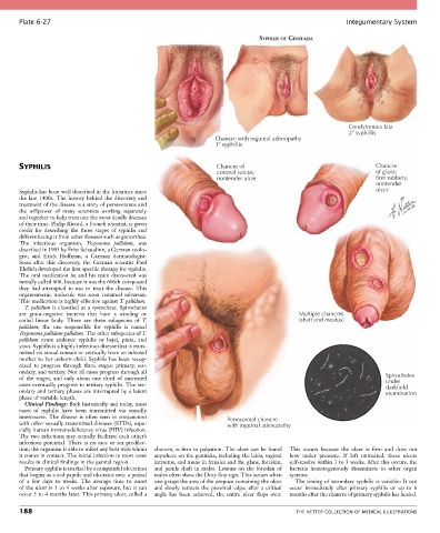
Plate 6-27                                                                                            Integumentary System

                                                                                  SYPHILIS OF GENITALIA


                                                                                                             Condylomata lata
                                                                                                             2  syphillis
                                                                    Chancre with inguinal adenopathy
                                                                    1  syphillis


       SYPHILIS                                                     Chancre of                                        Chancre
                                                                    coronal sulcus;                                   of glans:
                                                                    nontender ulcer                                   firm rubbery,
                                                                                                                      nontender
       Syphilis has been well described in the literature since                                                       ulcer
       the late 1400s. The history behind the discovery and
       treatment of the disease is a story of perseverance and
       the  willpower  of  many  scientists  working  separately
       and together to help treat one the most deadly diseases
       of their time. Philip Ricord, a French scientist, is given
       credit  for  describing  the  three  stages  of  syphilis  and
       differentiating it from other diseases such as gonorrhea.
       The  infectious  organism,  Treponema  pallidum,  was
       described in 1905 by Fritz Schaudinn, a German zoolo-
       gist,  and  Erich  Hoffman,  a  German  dermatologist.
       Soon  after  this  discovery,  the  German  scientist  Paul
       Ehrlich developed the first specific therapy for syphilis.
       The  oral  medication  he  and  his  team  discovered  was
       initially called 606, because it was the 606th compound
       they  had  attempted  to  use  to  treat  the  disease.  This
       organoarsenic  molecule  was  soon  renamed  salvarsan.
       This medication is highly effective against T. pallidum.
         T. pallidum is classified as a spirochete. Spirochetes
       are  gram-negative  bacteria  that  have  a  winding  or                               Multiple chancres
       coiled  linear  body.  There  are  three  subspecies  of  T.                           (shaft and meatus)
       pallidum;  the  one  responsible  for  syphilis  is  named
       Treponema pallidum pallidum. The other subspecies of T.
       pallidum  cause  endemic  syphilis  or  bejel,  pinta,  and
       yaws. Syphilis is a highly infectious disease that is trans-
       mitted via sexual contact or vertically from an infected
       mother to her unborn child. Syphilis has been recog-
       nized  to  progress  through  three  stages:  primary,  sec-
       ondary, and tertiary. Not all cases progress through all
       of  the  stages,  and  only  about  one  third  of  untreated                                                     Spirochetes
                                                                                                                         under
       cases eventually progress to tertiary syphilis. The sec-                                                          darkfield
       ondary and tertiary phases are interrupted by a latent                                                            examination
       phase of variable length.
         Clinical Findings: Both historically and today, most
       cases  of  syphilis  have  been  transmitted  via  sexually
       intercourse.  The  disease  is  often  seen  in  conjunction     Penoscrotal chancre
       with other sexually transmitted diseases (STDs), espe-           with inguinal adenopathy
       cially human immunodeficiency virus (HIV) infection.
       The two infections may actually facilitate each other’s
       infectious potential. There is no race or sex predilec-
       tion; the organism is able to infect any host with whom   chancre, is firm to palpation. The ulcer can be found   This  occurs  because  the  ulcer  is  firm  and  does  not
       it comes in contact. The initial infection in most cases   anywhere on the genitalia, including the labia, vaginal   bow  under  pressure.  If  left  untreated,  these  ulcers
       results in clinical findings in the genital region.  introitus, and mons in females and the glans, foreskin,   self-resolve within 1 to 3 weeks. After this occurs, the
         Primary syphilis is marked by a nonpainful ulceration   and  penile  shaft  in  males.  Lesions  on  the  foreskin  of   bacteria  hematogenously  disseminate  to  other  organ
       that begins as a red papule and ulcerates over a period   males often show the Dory flop sign. This occurs when   systems.
       of  a  few  days  to  weeks.  The  average  time  to  onset     one grasps the area of the prepuce containing the ulcer   The timing of secondary syphilis is variable: It can
       of the ulcer is 3 to 4 weeks after exposure, but it can   and  slowly  retracts  the  proximal  edge;  after  a  critical   occur  immediately  after  primary  syphilis  or  up  to  6
       occur 3 to 4 months later. This primary ulcer, called a   angle  has  been  achieved,  the  entire  ulcer  flops  over.   months after the chancre of primary syphilis has healed.

       188                                                                                   THE NETTER COLLECTION OF MEDICAL ILLUSTRATIONS