
Page 75 - The Netter Collection of Medical Illustrations - Integumentary System_ Volume 4 ( PDFDrive )
P. 75
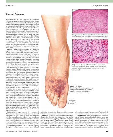
Plate 3-10                                                                                               Malignant Growths


        KAPOSI’S SARCOMA


        Kaposi’s  sarcoma  is  a  rare  malignancy  of  endothelial
        cells seen in unique settings. The classic variant is seen
        in older patients, most commonly individuals living in
        the region surrounding the Mediterranean Sea. Kaposi’s
        sarcoma associated with human immunodeficiency virus
        (HIV)  infection  or  with  acquired  immunodeficiency
        syndrome  (AIDS)  is  seen  predominantly  in  men,  and
        the tumor is thought to be caused by human herpesvirus-8
        (HHV8).  There  is  also  a  variant  seen  in  chronically
        immunosuppressed  patients,  such  as  those  who  have                         Low power. An abnormal proliferation of blood vessels
        undergone  solid  organ  transplantation.  The  African                         with slitlike spaces and extravasation of red blood cells
        cutaneous variant of Kaposi’s sarcoma is seen in younger
        men  in  their  third  or  fourth  decade  of  life.  Kaposi’s
        sarcoma is a locally aggressive tumor that rarely has a
        fatal  outcome.  The  one  exception  is  the  very  rare
        African  lymphadenopathic  form  of  Kaposi’s  sarcoma,
        which is distinct from the more common African cuta-
        neous form.
          Clinical  Findings:  The  tumors  are  very  similar  in
        appearance across the subtypes of clinical settings. They
        usually appear as pink-red to purple macules, papules,
        plaques,  or  nodules.  In  the  classic  form  of  Kaposi’s
        sarcoma, the tumors are most often found on the lower
        extremities of older men. Some tumors in this setting
        remain unchanged for years, and the patient often dies
        of  other  causes.  Occasionally,  the  tumors  grow  and
        ulcerate, causing pain and bleeding. The disseminated
        form of classic Kaposi’s sarcoma can be very aggressive,                        High power. Plump endothelial cells, with mutiple
        and patients require systemic chemotherapy.                                     abnormal-appearing blood vessels in a disorganized
          AIDS-associated  Kaposi’s  sarcoma  is  the  most
        common form of the disease. It is most often seen in                            pattern. Multiple extravasated red blood cells are
                                                                                        appreciated.
        younger men. In comparison with the classic form, this
        form usually manifests as purple macules, plaques, and
        nodules on the head and neck, trunk, and upper extrem-
        ities.  This  is  an  AIDS-defining  illness.  Patients  with
        AIDS-associated Kaposi’s sarcoma are at a higher risk
        for  internal  organ  involvement.  The  small  bowel  has
        been reported to be the internal organ most commonly
        affected by Kaposi’s sarcoma, but it can affect any organ
        system. Since the advent of multiple-drug therapy for
        HIV infection, the incidence of AIDS-associated Kapo-                         Kaposi’s sarcoma.
        si’s sarcoma has decreased dramatically.                                      Classic Kaposi’s sarcoma presenting
          Tropical African cutaneous Kaposi’s sarcoma is most                         on the lower extremity as purplish
        often seen in younger men. The clinical findings are not                      papules, plaques, and nodules
        much different from those of the classic form of Kapo-
        si’s  sarcoma.  These  patients  are  much  more  likely  to
        suffer from severe lower-extremity edema. The tumor
        also has a higher incidence of bone invasion than the
        other  types.  The  main  difference  between  the  classic
        and the African forms of Kaposi’s sarcoma is the age at
        onset. The aggressive form of African Kaposi’s sarcoma
        occurs  in  childhood  and  is  often  fatal  because  of  its
        aggressive ability to metastasize. The lymph nodes are
        often  involved  before  the  skin  is.  The  reason  the
        African  forms  act  so  differently  from  each  other  is
        poorly understood.
          Pathogenesis:  The  pathogenesis  of  the  classic  and   endothelial cells, allowing them to proliferate uncon-  of vascular spaces and a large amount of red blood cell
        African forms of Kaposi’s sarcoma is unknown. The cell   trolled by normal immune functions.  extravasation into the dermis.
        of origin of this tumor is believed to be the endothelial   Histology: Biopsies of Kaposi’s sarcoma show many   Treatment: For classic Kaposi’s sarcoma, the main-
        cell. Matrix metalloproteinases 2 and 9 have been shown   characteristic  findings.  The  promontory  sign  is  often   stay of therapy has been localized radiation treatment.
        to increase angiogenesis and increase the tissue invasion   seen; it is represented by plump endothelial cells jutting   Many other treatments have been advocated, including
        of the affected endothelial cells. Kaposi’s sarcoma asso-  into  the  lumen  of  the  capillary  vessel.  Many  slit-like   topical  alitretinoin,  imiquimod,  intralesional  vincris-
        ciated with AIDS or other immunosuppressive states is   spaces  are  also  seen.  These  spaces  represent  poorly   tine,  and  interferon.  Systemic  chemotherapy  for  dis-
        believed to be caused by the action of HHV8 in a geneti-  formed blood vessels, which are thin walled and easily   seminated  and  aggressive  forms  is  indicated  and  is
        cally predisposed individual. HHV8 is thought to cause   compressed. They are filled with red blood cells. The   usually based on a regimen of either vinblastine, pacli-
        dysregulation of the immune response in the afflicted   tumor in general is very vascular, with a predominance   taxel, bleomycin, or pegylated liposomal doxorubicin.


        THE NETTER COLLECTION OF MEDICAL ILLUSTRATIONS                                                                           61