
Page 80 - The Netter Collection of Medical Illustrations - Integumentary System_ Volume 4 ( PDFDrive )
P. 80
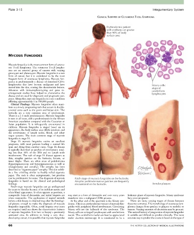
Plate 3-15                                                                                            Integumentary System

                                                                      CLINICAL SUBTYPES OF CUTANEOUS T-CELL LYMPHOMA


                                                                            Erythrodermic patient
                                                                            with erythema on greater
                                                                            than 90% of body
                                                                            surface area


       MYCOSIS FUNGOIDES


       Mycosis fungoides is the most common form of cutane-
       ous T-cell lymphoma. The cutaneous T-cell lympho-
       mas  are  an  assorted  group  of  cancers  with  varying
       genotypes and phenotypes. Mycosis fungoides is a rare
       form  of  cancer,  but  it  is  considered  to  be  the  most
       frequent  form  of  cutaneous  lymphoma.  Mycosis  fun-
       goides is predominantly a disease of abnormal CD4+
       lymphocytes  that  have  become  malignant  and  have                                                Sézary cells:
       moved into the skin, causing the characteristic lesions.                                             atypical
       Advances  with  immunophenotyping  and  gene  re-                                                    cerebriform
       arrangement  studies  have  helped  to  characterize  the                                            lymphocytes
       disease and are used for diagnostic and prognostic pur-
       poses. Altogether, mycosis fungoides is a rare condition
       afflicting approximately 1 in 500,000 people.
         Clinical  Findings:  Mycosis  fungoides  often  mani-
       fests as a slowly progressing rash that occurs in double-
       covered  areas  such  as  the  groin  and  breast  skin.  The
       buttocks  are  a  very  common  area  of  involvement.
       There is a 2 : 1 male predominance. Mycosis fungoides
       is seen in all races, with a predominance in the African
       American population compared with the Caucasian or
       Asian  population.  It  is  infrequently  encountered  in
       children.  Mycosis  fungoides  is  staged  based  on  its
       appearance, the body surface area (BSA) involved, and
       the  involvement  of  lymph  nodes,  blood,  and  other
       organ  systems.  The  most  common  stage  of  mycosis
       fungoides is stage IA.
         Stage  IA  mycosis  fungoides  carries  an  excellent
       prognosis,  with  most  patients  leading  a  normal  life
       span  and  dying  from  another  cause.  Stage  IA  disease
       is typically described as patches of involvement total-
       ing  less  than  10%  of  the  BSA  and  no  lymph  node
       involvement.  The  rash  of  stage  IA  disease  appears  as
       thin,  atrophic  patches  on  the  buttocks,  breasts,  or
       inner  thighs.  There  are  often  areas  of  poikiloderma
       (hyperpigmentation  and  hypopigmentation  as  well  as
       telangiectasias  and  atrophy).  The  atrophy  has  been
       described as “cigarette paper” atrophy: The skin exhib-
       its  a  fine  crinkling  similar  to  freshly  rolled  cigarette
       paper.  The  rash  is  often  asymptomatic,  but  pruritus
       can be problematic for some. The diagnosis of mycosis   Patch stage of mycosis fungoides on the buttocks.
       fungoides  is  based  on  the  clinical  and  pathological   Atrophic poikilodermatous patches are frequently
       findings.                                      encountered on the buttocks.                          Annular plaques
         Patch-stage  mycosis  fungoides  can  go  undiagnosed
       for years to decades because of its indolent nature and
       often bland appearance. It often appears as psoriasis, a
       nonspecific form of dermatitis, and initial biopsies are   may start as a form of dermatitis and over many years   leukemic phase of mycosis fungoides. Sézary syndrome
       often  nonspecific.  The  application  of  topical  steroids   transform into a malignant CD4+ process.  has a poor prognosis.
       before a skin biopsy is obtained may alter the histologi-  At the other end of the spectrum is the Sézary syn-  There  are  many  varying  stages  of  disease  between
       cal  picture  enough  to  make  the  diagnosis  of  mycosis   drome. This is an erythrodermic variant of mycosis fun-  these two extremes. The morphology of cutaneous lym-
       fungoides impossible. Often, serial biopsies over years   goides with peripheral blood involvement. Circulating   phoma changes from patches to plaques to nodules or
       are required until one shows the characteristic features   Sézary  cells  are  the  hallmark  of  this  syndrome.  The   tumors. Varying amounts of ulceration may be present.
       of mycosis fungoides. It is best to biopsy a previously   Sézary cells are enlarged lymphocytes with cerebriform   The natural history of progression of mycosis fungoides
       untreated  area.  In  addition  to  being  a  very  slow-  nuclei. The cerebriform nuclei can best be appreciated   is variable and difficult to predict clinically. The most
       developing cancer, it is possible that mycosis fungoides   under  electron  microscopy.  It  is  considered  to  be  a   accurate way to predict the course is based on the type of

       66                                                                                    THE NETTER COLLECTION OF MEDICAL ILLUSTRATIONS