
Page 82 - The Netter Collection of Medical Illustrations - Integumentary System_ Volume 4 ( PDFDrive )
P. 82
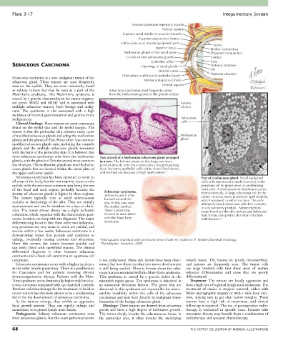
Plate 3-17                                                                                            Integumentary System

                                                                 Levator palpebrae superioris muscle
                                                                                Orbital septum
                                                            Superior tarsal (Müller’s) muscle (smooth)
                                                                    Superior conjunctival fornix
                                                            Orbicularis oculi muscle (palpebral part)   Sclera
                                                                            Superior tarsus            Bulbar conjunctiva
                                                             Meibomian glands of the tarsal plate      Palpebral conjunctiva
                                                              Glands of Zeis (sebaceous glands)        Cornea
                                                                          Eyelashes (cilia)            Lens
       SEBACEOUS CARCINOMA                                           Openings of tarsal glands         Anterior chamber
                                                                              Inferior tarsus          Iris
                                                            Orbicularis oculi muscle (palpebral part)  Posterior chamber
       Sebaceous carcinoma is a rare malignant tumor of the
       sebaceous  gland.  These  tumors  are  most  frequently         Inferior conjunctival fornix
       seen on the eyelids. They are most commonly found                       Orbital septum
       as  solitary  tumors  but  may  be  seen  as  a  part  of  the   Sebaceous carcinoma most frequently arises
       Muir-Torre  syndrome.  The  Muir-Torre  syndrome  is   from the meibomian glands or the glands of Zeis.
       caused by a genetic abnormality in the tumor suppres-
       sor  genes  MSH2  and  MLH1  and  is  associated  with                           Lumen
       multiple  sebaceous  tumors,  both  benign  and  malig-                          of duct
       nant.  The  syndrome  is  also  associated  with  a  high
       incidence of internal gastrointestinal and genitourinary
       malignancies.                                                                     Sebaceous
         Clinical Findings: These tumors are most commonly                               cell
       found  on  the  eyelid  skin  and  the  eyelid  margin.  The
       reason is that the periocular skin contains many types
       of modified sebaceous glands, including the meibomian                            Meibomian
       glands and the glands of Zeis. Many other, less common                           gland
       modified sebaceous glands exist, including the caruncle
       glands  and  the  multiple  sebaceous  glands  associated
       with the hairs of the periocular skin. It is believed that
       most sebaceous carcinomas arise from the meibomian   Two alveoli of a Meibomian sebaceous gland arranged
       glands, with the glands of Zeis the second most common   in a row. The left one seems to discharge secretory
       site of origin. The meibomian glands are modified seba-  product directly onto the surface into a straight opening
       ceous glands that are located within the tarsal plate of   duct. Secretory epithelial cells of the alveoli look foamy
       the upper and lower eyelid.               and washed out because of high lipid content.*
         Sebaceous carcinoma has been reported to occur in                                        Part of a sebaceous gland. Small nucleated
       all areas of the body, but the vast majority occur on the                                  cells with euchromatic nuclei (arrows) in the
       eyelids, with the next most common area being the rest                                     periphery of the gland serve as proliferating
       of  the  head  and  neck  region,  probably  because  the                                  stem cells. A thin basement membrane covers
       density of sebaceous glands is higher in these regions.   Sebaceous carcinoma.             them externally. A large sebaceous cell in the
                                                   Yellowish patch often
       The  tumors  typically  start  as  small  subcutaneous   located around the                center contains many prominent lipid droplets,
       nodules  or  thickenings  of  the  skin.  They  are  initially   eye, in this case near    which surround a central nucleus. The cells
       asymptomatic and can be mistaken for a stye or chala-  the medial canthus.                 ultimately break down and add their contents
                                                                                                  to oily secretory product.  Sebum reduces
       zion. The tumor almost always has a slight yellowish   These tumors may                    water loss from the skin surface and lubricates
       coloration, which, together with the characteristic peri-  be seen in association          hair. It may also protect skin from infection
       ocular location, can help with the diagnosis. The major   with the Muir-Torre              with bacteria.*
       differentiating factor is that these other two inflamma-  Syndrome.
       tory processes are very acute in onset, are painful, and
       resolve within a few weeks. Sebaceous carcinoma is a
       slow-growing  tumor  that  persists  and  continues  to
       enlarge,  eventually  causing  erosions  and  ulceration.   *Micrographs reprinted with permission from Ovalle W, Nahirney P. Netter’s Essential Histology.
       Once  this  occurs,  the  tumor  becomes  painful  and      Philadelphia: Saunders, 2008.
       can  easily  bleed  with  superficial  trauma.  The  clinical
       differential  diagnosis  is  often  between  sebaceous
       carcinoma and a basal cell carcinoma or squamous cell
       carcinoma.                                is not understood. Many risk factors have been deter-  muscle  tissue.  The  lesions  are  poorly  circumscribed,
         Sebaceous carcinomas occur with a higher incidence   mined, but how these translate into tumor development   and  mitoses  are  frequently  seen.  The  tumor  cells
       in the older female population. There is a predilection   is still being studied. More is known about the seba-  are  large  basaloid  cells  that  show  areas  of  mature
       for  Caucasians  and  for  patients  receiving  chronic   ceous tumors associated with the Muir-Torre syndrome.   sebocyte  differentiation  and  areas  that  are  poorly
       immunosuppressive  therapy.  Patients  with  the  Muir-  This  syndrome  is  caused  by  a  genetic  defect  in  the   differentiated.
       Torre syndrome are at dramatically higher risk for seba-  mismatch repair genes. The syndrome is inherited in   Treatment:  The  tumors  are  locally  aggressive  and
       ceous carcinoma compared with age-matched controls.   an  autosomal  dominant  fashion.  The  genes  that  are   have a high rate of regional lymph node metastasis. The
       Previous radiation therapy for the treatment of facial or   abnormal in this syndrome are responsible for micro-  treatment  of  choice  is  surgical  removal,  either  with
       ocular tumors has also been shown to be a predisposing   satellite  instability  within  the  cells  of  the  sebaceous   Mohs micrographic surgery or with a wide local exci-
       factor for the development of sebaceous carcinoma.  carcinomas and may lead directly to malignant trans-  sion,  making  sure  to  get  clear  tumor  margins.  These
         As  the  tumors  enlarge,  they  exhibit  an  aggressive   formation of the benign sebaceous gland.  tumors  have  a  high  risk  of  recurrence,  and  clinical
       local  growth  pattern.  They  can  rapidly  enlarge  and   Histology: These tumors are derived from sebaceous   follow-up is required. The use of postoperative radio-
       metastasize to regional lymph node basins.  glands and show a high degree of infiltrative growth.   therapy  is  warranted  in  specific  cases.  Patients  with
         Pathogenesis:  Solitary  sebaceous  carcinomas  arise   The tumor deeply invades the subcutaneous tissue; in   metastatic disease may benefit from a combination of
       from sebaceous glands, but the exact pathomechanism   the  periocular  area,  it  often  invades  the  underlying   radiotherapy and systemic chemotherapy.

       68                                                                                    THE NETTER COLLECTION OF MEDICAL ILLUSTRATIONS