
Page 81 - The Netter Collection of Medical Illustrations - Integumentary System_ Volume 4 ( PDFDrive )
P. 81
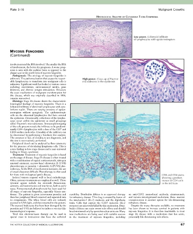
Plate 3-16                                                                                               Malignant Growths

                                                                     HISTOLOGICAL ANALYSIS OF CUTANEOUS T-CELL LYMPHOMA


                                                                                                Low power. Lichenoid infiltrate
                                                                                                of lymphocytes with epidermotropism


        MYCOSIS FUNGOIDES
        (Continued)


        involvement and the BSA involved. The smaller the BSA
        of involvement, the better the prognosis. A worse prog-
        nosis is seen with the nodular form as opposed to the
        plaque type or the patch form of mycosis fungoides.
          Pathogenesis: The etiology of mycosis fungoides is
        unknown. The pathomechanism that causes the respon-  High power. Close-up of Pautrier
        sible lymphocytes to transform into malignant cells is   microabscess in the epidermis
        unknown. Significant work has looked at various causes
        including  retroviruses,  environmental  insults,  gene
        deletions,  and  chronic  antigen  stimulation.  However,
        the exact mechanism of malignant transformation for
        this  disease,  which  was  originally  described  in  1806,
        remains unresolved.
          Histology: Stage IA disease shows the characteristic
        histological findings of mycosis fungoides. There is a
        lichenoid infiltrate of abnormal lymphocytes with cere-
        briform nuclei. There are varying amounts of epider-
        motropism  without  spongiosis.  The  epidermotropic
        cells are the abnormal lymphocytes that have entered
        the epidermis. Occasionally, collections of the lympho-
        cytes  occur  within  the  epidermis  as  small  groupings
        called  Pautrier’s  microabscesses.  Immunophenotyping
        of the cells present reveals the infiltrate to be predomi-
        nantly CD4+ lymphocytes with a loss of the CD7 and
        CD26 surface molecules. Clonality of the infiltrate can
        be determined by performing a Southern blot analysis.
        The presence or lack of clonality is not diagnostic, and
        this test is not routinely performed.
          Peripheral blood can be analyzed by flow cytometry
        for the presence of circulating lymphoma cells. This is
        a rare finding in low-stage disease and a near-universal
        finding in Sézary syndrome.
          Treatment: Treatment of mycosis fungoides is based
        on the stage of disease. Stage IA disease is often treated
        with a combination of topical corticosteroids, nitrogen
        mustard  ointment,  narrow-band  ultraviolet  B  (UVB)
        phototherapy, or psoralen + ultraviolet A (PUVA) pho-
        totherapy. As the BSA of involvement increases, the use
        of creams becomes difficult. Phototherapy is often used
        for those with widespread patch disease.                                                                    CD8 and CD4 stains
          Isolated tumors respond well to local radiotherapy.                                                       showing a predom-
        Often, systemic treatments are employed as well. These   CD8                 CD4                            inance of CD4 cells
        systemic  agents  include  the  retinoids  (bexarotene,                                                     in the infiltrate
        acitretin, and isotretinoin) and interferon, both α and γ
        types. Extracorporeal photophoresis has been used for
        all stages of mycosis fungoides, especially Sézary syn-
        drome. The patient is given intravenous psoralen and   capability.  Denileukin  diftitox  is  an  approved  therapy   an  anti-CD52  monoclonal  antibody,  alemtuzumab,
        then has peripheral blood removed and separated into   for refractory disease. This drug is created by fusion of   and  various  investigational  mediations.  Bone  marrow
        its  components.  The  white  blood  cells  are  isolated,   the  interleukin-2  (IL-2)  molecule  and  the  diphtheria   transplantation  is  another  option  for  life-threatening
        exposed to UVA light, and then returned to the patient.   toxin.  Cells  that  express  the  CD25  molecule  (IL-2   refractory disease.
        The exposed leukocytes that have been damaged by the   receptor) are selectively killed by this medication. Deni-  Despite the many therapies available, no treatment
        psoralen and UVA are believed to induce a vaccine-like   leukin diftitox can cause severe side effects and should   has  been  shown  to  increase  survival  in  patients  with
        immunological response.                   be administered only by specialists adept at its use. Many   mycosis  fungoides.  It  is  therefore  inadvisable  to  treat
          Total  skin  electron-beam  therapy  can  be  used  in   new medications are being used with variable success     stage  IA  disease  with  a  medication  that  has  acute,
        special  cases  in  institutions  that  have  the  technical   in  the  treatment  of  mycosis  fungoides,  including     potentially life-threatening side effects.


        THE NETTER COLLECTION OF MEDICAL ILLUSTRATIONS                                                                           67