
Page 78 - The Netter Collection of Medical Illustrations - Integumentary System_ Volume 4 ( PDFDrive )
P. 78
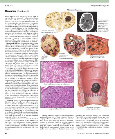
Plate 3-13                                                                                            Integumentary System

       MELANOMA (Continued)                                                      METASTATIC MELANOMA


       slowly  enlarging  pink  patches  or  plaques  with  no
       pigment. They are commonly misdiagnosed as derma-
       titis  or  tinea  infections,  and  the  diagnosis  is  often                                                 CT with contrast
       delayed. They can also resemble actinic keratoses. The                                                        enhancement
       lack of pigment takes away the clinician’s most impor-                                                        shows a similar
       tant diagnostic clue. These tumors are often biopsied                                                         large metastasis
       because they have not gone away after being treated for                                                       in the right
       something entirely different or after they have devel-                                                        cerebellum with
       oped a papule or nodule. At that point, they are still                                                        effacement of the
       most commonly thought to be basal cell carcinomas or   Cerebellar metastasis                                  fourth ventricle
       squamous  cell  carcinomas;  rarely  does  the  clinician   from cutaneous melanoma
       include amelanotic melanoma in the differential diag-
       nosis. Patients with albinism or xeroderma pigmento-
       sum are at a higher risk for development of amelanotic
       melanoma.  These  patients  need  to  be  screened  rou-
       tinely, and any suspicious lesions should be biopsied.
         Pathogenesis: There is no single gene defect that can
       explain the development of all melanomas. The most
       plausible theory is that a melanocyte within the epider-
       mis is damaged by some external event, such as chronic
       ultraviolet exposure, or by some internal event, such as
       the spontaneous mutation of a key gene in the regula-
       tion of cell proliferation or apoptosis. After this event       Multiple
       has  occurred,  the  abnormal  melanocyte  proliferates         metastases to
       with  the  epidermis,  starting  as  an  in  situ  variant  of   heart from                          Malignant melanoma
       melanoma. After time, the clonal melanoma cells begin           malignant melanoma                   metastases to the liver
       to  coalesce  and  form  nests  of  melanoma  cells.  They
       then continue to proliferate and enlarge until the clini-
       cal  features  are  evident.  The  tumor  enters  a  radial
       growth phase at first and eventually develops a vertical
       growth phase with metastatic potential.
         Approximately 10% of melanomas are considered to
       be  an  inherited  familial  form.  Although  no  one  gene
       explains all of these tumors, the p16 gene (TP16) is likely
       the main susceptibility gene. This gene, when mutated,
       increases  an  individual’s  risk  for  melanoma  as  well  as
       pancreatic carcinoma. TP16 is a tumor suppressor gene
       that  is  inherited  in  an  autosomal  dominant  fashion.
       Genetic testing for this gene is commercially available.
         Histology: The diagnosis by histology of melanoma
       is based on multiple criteria, including symmetry, mela-  Sheets of bizarre-appearing melanocytes
       nocyte atypia, mitosis, distribution of the melanocytes
       within the epidermis, lack of maturation of melanocytes
       as they extend deeper into the dermis, circumscription,
       and  architectural  disorder.  Melanoma  is  believed  to
       begin with an in situ portion, followed by an upward
       spread  of  single  melanocytes  within  the  epidermis,
       termed pagetoid spread. If no epidermal component of
       melanoma is seen, the possibility of a metastatic focus
       is entertained.
         Treatment: When a clinician encounters a pigmented
       skin lesion that is believed to be a melanoma, the lesion
       should  be  biopsied  promptly.  The  best  method  for
       biopsy of a pigmented lesion that is suspicious for mela-
       noma is with an excisional biopsy method using a small                                           Melanoma metastasis
       (1-2 mm)  margin  of  normal  surrounding  skin.  This   Large nodular melanoma                  to the large intestine
       allows for the diagnosis and an accurate measurement
       of the Breslow depth. The Breslow depth is the distance
       from the granular cell layer to the base of the tumor.
       This  depth  is  considered  to  be  the  most  important   Sentinel lymph node sampling is becoming routinely   dissection  and  adjunctive  therapy  with  interferon.
       prognostic indicator for melanoma.        performed  in  the  care  of  these  patients  and  aids  in   Those  with  widespread  metastatic  disease  are  given
         Therapy for melanoma is based on the Breslow thick-  staging of the disease. If the patient has a positive sen-  various  chemotherapeutic  regimens  or  enrolled  into
       ness, the presence of ulceration, and the mitotic rate of   tinel  lymph  node  biopsy  for  metastatic  melanoma,   clinical studies. The mortality rate for stage IV mela-
       the primary tumor. The standard of care is to perform   staging  is  performed  based  on  positron  emission   noma is very poor. Follow-up for melanoma patients is
       a wide local excision with varying margins of skin based   tomography/computed  tomography  (PET/CT)  scan-  based on the stage of disease. The National Compre-
       on the criteria described previously. Melanoma in situ   ning  and  magnetic  resonance  imaging  (MRI)  of  the   hensive  Cancer  Network/National  Cancer  Institute
       is treated surgically by wide local excision with 5-mm   brain. Patients with metastatic disease to local lymph   (NCCN/NCI)  has  published  standardized  guidelines
       margins.                                  nodes  only  are  offered  a  localized  lymph  node   for clinicians.

       64                                                                                    THE NETTER COLLECTION OF MEDICAL ILLUSTRATIONS