
Page 77 - The Netter Collection of Medical Illustrations - Integumentary System_ Volume 4 ( PDFDrive )
P. 77
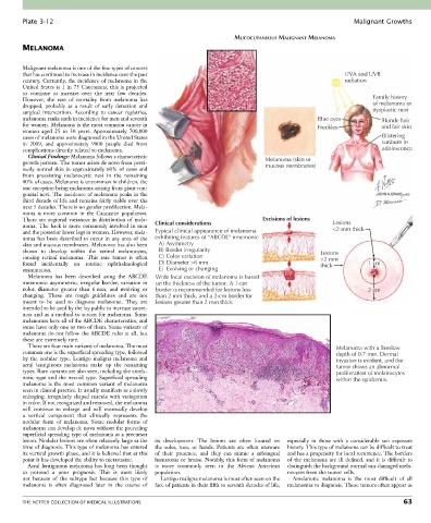
Plate 3-12                                                                                               Malignant Growths

                                                                           MUCOCUTANEOUS MALIGNANT MELANOMA
        MELANOMA

                                                                                                              Risk factors include:
        Malignant melanoma is one of the few types of cancers
        that has continued to increase in incidence over the past                                             UVA and UVB
        century. Currently, the incidence of melanoma in the                                                  radiation
        United  States  is  1  in  75  Caucasians;  this  is  projected
        to  continue  to  increase  over  the  next  few  decades.
        However,  the  rate  of  mortality  from  melanoma  has                                                        Family history
        dropped,  probably  as  a  result  of  early  detection  and                                                   of melanoma or
        surgical  intervention.  According  to  cancer  registries,                                                    dysplastic nevi
        melanoma ranks sixth in incidence for men and seventh                                         Blue eyes           Blonde hair
        for women. Melanoma is the most common cancer in                                              Freckles            and fair skin
        women  aged  25  to  30  years.  Approximately  700,000
        cases of melanoma were diagnosed in the United States                                                             Blistering
        in  2009,  and  approximately  9000  people  died  from                                                           sunburn in
        complications directly related to melanoma.                                                                       adolescence
          Clinical Findings: Melanoma follows a characteristic
        growth pattern. The tumor arises de novo from previ-                         Melanoma (skin or
                                                                                     mucous membranes)
        ously normal skin in approximately 60% of cases and
        from  preexisting  melanocytic  nevi  in  the  remaining
        40% of cases. Melanoma is uncommon in children, the
        one exception being melanoma arising from giant con-
        genital nevi. The incidence of melanoma peaks in the
        third decade of life and remains fairly stable over the
        next 5 decades. There is no gender predilection. Mela-
        noma  is  more  common  in  the  Caucasian  population.
        There  are  regional  variances  in  distribution  of  mela-                Excisions of lesions   Lesions
        noma.  The  back  is  more  commonly  involved  in  men   Clinical considerations                  <2 mm thick
        and the posterior lower legs in women. However, mela-  Typical clinical appearance of melanoma
        noma has been described to occur in any area of the   exhibiting features of “ABCDE” mnemonic
        skin and mucous membranes. Melanoma has also been   A)  Asymmetry
        shown  to  develop  within  the  retinal  melanocytes,   B)  Border irregularity               Lesions
        causing  retinal  melanoma.  This  rare  tumor  is  often   C)  Color variation                >2 mm
        found  incidentally  on  routine  ophthalmological   D) Diameter >6 mm                         thick          1 cm
        examination.                               E)  Evolving or changing
          Melanoma  has  been  described  using  the  ABCDE   Wide local excision of melanoma is based
        mnemonic: asymmetric, irregular border, variation in   on the thickness of the tumor. A 1-cm
        color,  diameter  greater  than  6 mm,  and  evolving  or   border is recommended for lesions less            2 cm
        changing.  These  are  rough  guidelines  and  are  not   than 2 mm thick, and a 2-cm border for
        meant  to  be  used  to  diagnose  melanoma.  They  are   lesions greater than 2 mm thick.
        intended to be used by the lay public to increase aware-
        ness and as a method to screen for melanoma. Some
        melanomas have all of the ABCDE characteristics, and
        some have only one or two of them. Some variants of
        melanoma do not follow the ABCDE rules at all, but
        these are extremely rare.
          There are four main variants of melanoma. The most                                                Melanoma with a Breslow
        common one is the superficial spreading type, followed                                              depth of 0.7 mm. Dermal
        by  the  nodular  type.  Lentigo  maligna  melanoma  and                                            invasion is evident, and the
        acral  lentiginous  melanoma  make  up  the  remaining                                              tumor shows an abnormal
        types. Rare variants are also seen, including the amela-                                            proliferation of melanocytes
        notic  type  and  the  nevoid  type.  Superficial  spreading                                        within the epidermis.
        melanoma  is  the  most  common  variant  of  melanoma
        seen in clinical practice. It usually manifests as a slowly
        enlarging,  irregularly  shaped  macule  with  variegation
        in color. If not recognized and removed, the melanoma
        will  continue  to  enlarge  and  will  eventually  develop
        a  vertical  component  that  clinically  represents  the
        nodular  form  of  melanoma.  Some  nodular  forms  of
        melanoma can develop de novo without the preceding
        superficial spreading type of melanoma as a precursor
        lesion. Nodular lesions are often relatively large at the   its  development.  The  lesions  are  often  located  on    especially  in  those  with  a  considerable  sun  exposure
        time of diagnosis. This type of melanoma has entered   the  soles,  toes,  or  hands.  Patients  are  often  unaware     history. This type of melanoma can be difficult to treat
        its vertical growth phase, and it is believed that at this   of  their  presence,  and  they  can  mimic  a  subungual   and has a propensity for local recurrence. The borders
        point it has developed the ability to metastasize.  hematoma or bruise. Notably, this form of melanoma   of  the  melanoma  are  ill  defined,  and  it  is  difficult  to
          Acral lentiginous melanoma has long been thought   is  more  commonly  seen  in  the  African  American   distinguish the background normal sun-damaged mela-
        to  portend  a  poor  prognosis.  This  is  most  likely     population.            nocytes from the tumor cells.
        not  because  of  the  subtype  but  because  this  type  of   Lentigo maligna melanoma is most often seen on the   Amelanotic  melanoma  is  the  most  difficult  of  all
        melanoma  is  often  diagnosed  later  in  the  course  of     face of patients in their fifth to seventh decades of life,   melanomas to diagnosis. These tumors often appear as


        THE NETTER COLLECTION OF MEDICAL ILLUSTRATIONS                                                                           63