
Page 83 - The Netter Collection of Medical Illustrations - Integumentary System_ Volume 4 ( PDFDrive )
P. 83
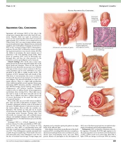
Plate 3-18                                                                                               Malignant Growths

                                                                              GENITAL SQUAMOUS CELL CARCINOMA


                                                                                                        Extensive
                                                                                                        fungating
                                                                                                        carcinoma
                                                                                                        of penis


        SQUAMOUS CELL CARCINOMA


        Squamous  cell  carcinoma  (SCC)  of  the  skin  is  the
        second most common skin cancer after basal cell carci-
        noma.  Together,  these  two  types  of  carcinoma  are
        known as non-melanoma skin cancer. SCC accounts for
        approximately 20% of all skin cancers diagnosed in the
        United States. SCC can come in many variants, includ-
        ing in situ and invasive types. Bowen’s disease, bowenoid                      Extensive involvement
        papulosis, and erythroplasia of Queyrat are all forms of                       of presymphysial
        SCC in situ. A unique subtype of SCC is the keratoac-  Advanced carcinoma of penis  and inguinal nodes  Squamous cell carcinoma of
        anthoma. Invasive SCC is defined by invasion through                                               penis, histology
        the basement membrane zone into the dermis. SCC has
        the  ability  to  metastasize;  the  most  common  area  of
        metastasis  is  the  local  draining  lymph  nodes.  Most
        forms  of  cutaneous  SCC  occur  in  chronically  sun-
        damaged  skin,  and  they  are  often  preceded  by  the
        extremely common premalignant actinic keratosis.
          Clinical  Findings:  SCC  of  the  skin  is  most  com-
        monly located on the head and neck region and on the
        dorsal  hands  and  forearms.  These  are  the  areas  that
        obtain the most ultraviolet sun exposure over a lifetime.
        This type of skin cancer is more common in the Cau-
        casian population and in older individuals. It is more
        prevalent  in  the  fifth  to  eighth  decades  of  life.  The
        incidence  of  SCC  increases  with  each  decade  of  life.
        This  form  of  non-melanoma  skin  cancer  is  definitely
        linked to the amount of sun exposure one has had over
        one’s lifetime. Fair-skinned individuals are most com-
        monly  affected.  There  is  a  slight  male  predilection.
        Other risk factors include arsenic exposure, human pap-
        illomavirus  (HPV)  infection,  psoralen  +  ultraviolet  A
        light (PUVA) therapy, chronic scarring, chronic immu-  Erythroplasia of Queyrat            Carcinoma on leukoplakia
        nosuppression,  and  radiation  exposure.  Transplant
        recipients who are taking chronic immunosuppressive
        medications  often  develop  SCCs.  Their  skin  cancers
        also  tend  to  occur  on  the  head  and  neck  and  on  the
        arms,  but  in  addition  they  have  a  higher  percentage
        of  tumors  developing  on  the  trunk  and  other  non–
        sun-exposed regions.
          SCCs of the skin can occur with various morpholo-
        gies. They can start as thin patches or plaques. There
        is usually a thickened, adherent scale on the surface of
        the tumor. Variable amounts of ulceration are seen. As                  Early carcinoma
        the  tumors  enlarge,  they  can  take  on  a  nodular  con-
        figuration.  The  nodules  are  firm  and  can  be  deeply
        seated within the dermis. Most SCCs are derived from
        a  preexisting  actinic  keratosis.  Patients  often  have
        chronically  sun-damaged  skin  with  poikilodermatous                                Carcinoma under foreskin
        changes and multiple lentigines and actinic keratoses.
        Approximately 1% of actinic keratoses per year develop
        into SCC.
          Subungual  SCC  is  a  difficult  diagnosis  to  make
        without a biopsy. It is often preceded by an HPV infec-
        tion,  and  the  area  has  often  been  treated  for  long   diagnosis can be critical in sparing the patient an ampu-  SCC; two of the best recognized ones are epidermodys-
        periods  as  a  wart.  HPV  is  a  predisposing  factor,  and   tation of the affected digit.  plasia verruciformis and xeroderma pigmentosum.
        with time a small percentage of these warts transform   A few chronic dermatoses can predispose to the devel-  Pathogenesis: SCC is related to cumulative ultravio-
        into SCC. This development is usually associated with   opment of SCC, including lichen sclerosis et atrophicus,   let exposure. Ultraviolet B (UVB) light appears to be
        a subtle change in morphology. There tends to be more   disseminated and superficial actinic porokeratosis, warts,   the most important action spectrum in the development
        nail destruction and a slow enlargement over time in   discoid  lupus,  long-standing  ulcers,  and  scars.  Many   of SCC. UVB is much more potent than ultraviolet A
        the face of standard wart therapy. Prompt biopsy and   genetic diseases can predispose to the development of   light. UVB can damage keratinocyte DNA by causing


        THE NETTER COLLECTION OF MEDICAL ILLUSTRATIONS                                                                           69