
Page 76 - The Netter Collection of Medical Illustrations - Integumentary System_ Volume 4 ( PDFDrive )
P. 76
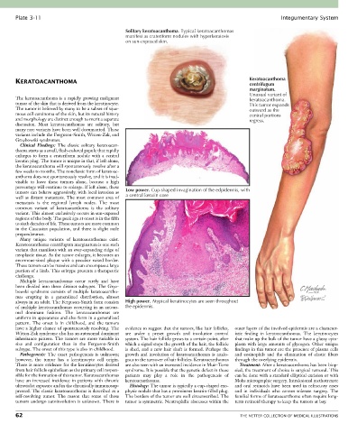
Plate 3-11                                                                                            Integumentary System

                                                 Solitary keratoacanthoma. Typical keratoacanthomas
                                                 manifest as crateriform nodules with hyperkeratosis
                                                 on sun-exposed skin.


                                                                                                 Keratoacanthoma
       KERATOACANTHOMA                                                                           centrifugum
                                                                                                 marginatum.
                                                                                                 Unusual variant of
       The  keratoacanthoma  is  a  rapidly  growing  malignant                                  keratoacanthoma.
       tumor of the skin that is derived from the keratinocyte.                                  This tumor expands
       The tumor is believed by many to be a subset of squa-                                     outward as the
       mous cell carcinoma of the skin, but its natural history                                  central portions
       and morphology are distinct enough to merit a separate                                    regress.
       discussion.  Most  keratoacanthomas  are  solitary,  but
       many rare variants have been well documented. These
       variants include the Ferguson-Smith, Witten-Zak, and
       Grzybowski syndromes.
         Clinical  Findings:  The  classic  solitary  keratoacan-
       thoma starts as a small, flesh-colored papule that rapidly
       enlarges  to  form  a  crateriform  nodule  with  a  central
       keratin plug. The tumor is unique in that, if left alone,
       the keratoacanthoma will spontaneously resolve after a
       few weeks to months. The nonclassic form of keratoac-
       anthoma does not spontaneously resolve, and it is inad-
       visable  to  leave  these  tumors  alone,  because  a  high
       percentage will continue to enlarge. If left alone, these
       tumors can behave aggressively, with local invasion as   Low power. Cup-shaped invagination of the edpidermis, with
       well as distant metastasis. The most common area of   a central keratin core
       metastasis  is  the  regional  lymph  nodes.  The  most
       common  variant  of  keratoacanthoma  is  the  solitary
       variant. This almost exclusively occurs in sun-exposed
       regions of the body. The peak age at onset is in the fifth
       to sixth decades of life. These tumors are more common
       in the Caucasian population, and there is slight male
       preponderance.
         Many  unique  variants  of  keratoacanthomas  exist.
       Keratoacanthoma centrifugum marginatum is one such
       variant that manifests with an ever-expanding ridge of
       neoplastic tissue. As the tumor enlarges, it becomes an
       enormous-sized  plaque  with  a  peculiar  raised  border.
       These tumors can be massive and can encompass a large
       portion of a limb. This subtype presents a therapeutic
       challenge.
         Multiple  keratoacanthomas  occur  rarely  and  have
       been  divided  into  three  distinct  subtypes.  The  Gryz-
       bowski  syndrome  consists  of  multiple  keratoacantho-
       mas  erupting  in  a  generalized  distribution,  almost
       always in an adult. The Ferguson-Smith form consists   High power. Atypical keratinocytes are seen throughout
       of multiple keratoacanthomas occurring in an autoso-  the epidermis.
       mal  dominant  fashion.  The  keratoacanthomas  are
       uniform in appearance and also form in a generalized
       pattern.  The  onset  is  in  childhood,  and  the  tumors
       have a higher chance of spontaneously resolving. The   evidence to suggest that the tumors, like hair follicles,   outer layers of the involved epidermis are a character-
       Witten-Zak syndrome also has an autosomal dominant   are  under  a  preset  growth  and  involution  control   istic  finding  in  keratoacanthomas.  The  keratinocytes
       inheritance pattern. The tumors are more variable in   system. The hair follicle grows to a certain point, after   that make up the bulk of the tumor have a glassy cyto-
       size  and  configuration  than  in  the  Ferguson-Smith   which a signal stops the growth of the hair, the follicle   plasm  with  large  amounts  of  glycogen.  Other  unique
       subtype. The onset of this type is also in childhood.  is  shed,  and  a  new  hair  shaft  is  formed.  Perhaps  the   findings in this tumor are the presence of plasma cells
         Pathogenesis:  The  exact  pathogenesis  is  unknown;   growth  and  involution  of  keratoacanthomas  is  analo-  and  eosinophils  and  the  elimination  of  elastic  fibers
       however,  the  tumor  has  a  keratinocyte  cell  origin.   gous to the turnover of hair follicles. Keratoacanthomas   through the overlying epidermis.
       There is more evidence for the keratinocytes derived   are also seen with an increased incidence in Muir-Torre   Treatment: After a keratoacanthoma has been biop-
       from hair follicle epithelium as the primary cell respon-  syndrome. It is possible that the genetic defect in these   sied, the treatment of choice is surgical removal. This
       sible for the formation of this tumor. Keratoacanthomas   patients  may  play  a  role  in  the  pathogenesis  of   can be done with a standard elliptical excision or with
       have  an  increased  incidence  in  patients  with  chronic   keratoacanthomas.     Mohs micrographic surgery. Intralesional methotrexate
       ultraviolet exposure and in the chronically immunosup-  Histology: The tumor is typically a cup-shaped exo-  and  oral  retinoids  have  been  used  in  refractory  cases
       pressed. The classic keratoacanthoma is described as a   phytic nodule that has a prominent keratin-filled plug.   and  in  individuals  who  cannot  tolerate  surgery.  The
       self-resolving  tumor.  The  reason  that  some  of  these   The borders of the tumor are well circumscribed. The   familial forms of keratoacanthoma often require long-
       tumors undergo autoinvolution is unknown. There is   tumor is symmetric. Neutrophilic abscesses within the   term retinoid therapy to keep the tumors at bay.

       62                                                                                    THE NETTER COLLECTION OF MEDICAL ILLUSTRATIONS