
Page 79 - The Netter Collection of Medical Illustrations - Integumentary System_ Volume 4 ( PDFDrive )
P. 79
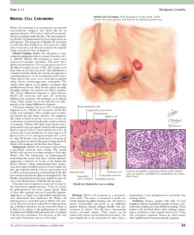
Plate 3-14                                                                                               Malignant Growths

                                                                   Merkel cell carcinoma. Pink-red papule on the cheek. These
        MERKEL CELL CARCINOMA                                      tumors may arise quickly and have an accelerated growth rate.


        Merkel cell carcinoma is an uncommonly encountered
        neuroendocrine  malignant  skin  tumor  that  has  an
        aggressive behavior. This tumor is derived from special-
        ized nerve endings within the skin. The tumor promot-
        ing Merkel cell polyomavirus has been implicated in its
        pathogenesis. The prognosis of Merkel cell carcinoma
        is worse than that of melanoma. This tumor has a high
        rate of recurrence and often has spread to the regional
        lymph nodes by the time of diagnosis.
          Clinical  Findings:  Merkel  cell  carcinoma  is  a  rare
        cutaneous malignancy with an estimated incidence of 1
        in  200,000.  Merkel  cell  carcinoma  is  much  more
        common  in  Caucasian  individuals.  The  tumor  has  a
        slight male predilection. The average age at onset is in
        the fifth to seventh decades of life. The lesions occur
        most often on the head and neck. This distribution is
        consistent with the notion that chronic sun exposure is
        a predisposing factor in the development of this tumor.
        These tumors also occur more commonly in patients
        taking  chronic  immunosuppressive  medications.  The
        tumors  often  appear  as  red  papules  or  plaques  that
        quickly increase in size. They can also appear as rapidly
        enlarging  nodules.  On  occasion,  the  tumor  ulcerates.
        The  clinical  differential  diagnosis  is  often  between
        Merkel  cell  carcinoma  and  basal  cell  carcinoma,
        inflamed cyst, squamous cell carcinoma, or an adnexal
        tumor.  These  tumors  are  so  rare  that  they  are  infre-
        quently in the original differential diagnosis.
          It has been estimated that up to 50% of all patients   Basal epithelial cells
        diagnosed  with  a  Merkel  cell  carcinoma  will  develop   Cytoplasmic protrusion
        lymph node metastasis. Other notable areas of metas-       Desmosomes
        tasis include the skin, lungs, and liver. The staging of           Merkel cell
        this tumor is based on its size (<2 cm or >2 cm), the
        involvement of regional lymph nodes, and the presence
        of metastasis. Patients with higher-stage disease have a
        progressively worse prognosis. Patients with metastatic
        disease (stage IV) have a 5-year survival rate of 0%. In
        contrast, the 5-year survival rate for local stage I or II
        disease is 65% to 75% and approximately 50% to 60%
        for stage III (lymph node involvement). Grouping all
        stages together, one third of the patients diagnosed with
        Merkel cell carcinoma will die from their disease.
          Pathogenesis: Merkel cell carcinoma is derived from
        a  specialized  cutaneous  nerve  ending.  The  normal
        Merkel cells function in mechanoreception of the skin.
        Merkel  cells,  like  melanocytes,  are  embryologically
        derived from the neural crest tissue. Chronic immuno-
        suppression  is  believed  to  be  one  of  the  largest  risk
        factors.  Patients  taking  immunosuppressive  medica-
        tions after organ transplantation are at much higher risk
        than age-matched controls. Chronic sun exposure and
        its effect on downregulating local immunity in the skin   Mitochondria  Schwann cell  Uniform basophilic-appearing Merkel cells. Merkel
                                                                                          cell carcinoma is classified as a small blue cell tumor.
        have also been theorized to play an etiological role. The   Expanded  Granulated vesicles  (H&E stain)
        Merkel cell polyomavirus has been studied to assess its   axon terminal  Lobulated nucleus
        role in the development of Merkel cell carcinoma.
          Polyomaviruses are similar in nature and structure to   Detail of a Merkel disc nerve ending
        the better-known papillomaviruses. There are at least
        five  polyomaviruses  that  cause  human  disease.  Most
        of  them  affect  patients  who  are  chronically  immuno-
        suppressed at a higher rate than healthy matched con-  Histology:  Merkel  cell  carcinoma  is  a  neuroendo-  characteristic,  if  not  pathognomonic  perinuclear  dot,
        trols.  Researchers  have  implicated  the  Merkel  cell   crine  tumor.  The  tumor  is  composed  of  small,  uni-  staining pattern.
        polyomavirus as a potential cause of Merkel cell carci-  formly shaped, basophilic-staining cells. The tumor is   Treatment:  Surgical  excision  with  wide  (2-3 cm)
        noma. This virus has been isolated from a high percent-  poorly  circumscribed  and  grows  in  an  infiltrative   margins is still the standard therapeutic treatment. Sen-
        age of Merkel cell tumors, but not from all of them. It   pattern  between  dermal  collagen  bundles  and  sub-  tinel node sampling has been helpful in staging. Those
        is likely to be a player in the pathogenesis of a subset   cutaneous  fat  lobules.  The  cells  have  a  characteristic   patients  with  localized  disease  often  undergo  postop-
        of patients with Merkel cell carcinoma, but it is unlikely   nuclear  chromatin  pattern.  These  tumors  can  be   erative irradiation of the surgical removal site. Those
        to be the only explanation. The discovery of this virus   stained with various immunohistochemical stains. The   with  widespread  metastatic  disease  are  often  treated
        may lead to therapeutic options in the future.  most  helpful  one  is  the  cytokeratin  20  stain.  It  has  a   with cisplatin-based chemotherapeutic regimens.


        THE NETTER COLLECTION OF MEDICAL ILLUSTRATIONS                                                                           65