
Page 89 - The Netter Collection of Medical Illustrations - Integumentary System_ Volume 4 ( PDFDrive )
P. 89
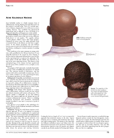
Plate 4-4                                                                                                            Rashes


        ACNE KELOIDALIS NUCHAE

        Acne  keloidalis  nuchae  is  a  fairly  common  form  of
        inflammatory, scarring alopecia that typically occurs on
        the posterior occipital scalp. There is a variable spec-
        trum of disease, ranging from very mild cases to severe
        scarring  alopecia.  The  condition  has  psychosocial
        implications  and  is  difficult  to  treat  effectively.  It  is
        diagnosed clinically, and biopsies are rarely needed.
          Clinical Findings: Acne keloidalis nuchae begins on
        the posterior scalp or nape of the neck as tiny, follicular,
        flesh-colored  to  red  papules.  The  papules  enlarge                              Mild. Follicle-centered,
        to  form  plaques,  which  coalesce  into  larger  plaques.                          flesh-colored papules
        Ultimately  in  severe  cases,  the  entire  posterior  scalp
        is involved. Early in the disease, no hair loss is appre-
        ciated.  As  the  disease  progresses,  the  hair  follicles
        become scarred down and crowded out by the encroach-
        ing fibrosis, resulting in a variable amount of scarring
        alopecia.
          This condition is far more common in young adult
        men, with a predilection for African Americans. It was
        originally  believed  to  be  caused  by  close  shaving  of
        the hair and the subsequent inflammation caused by the
        newly regrowing hair as it pierces the epidermis. The
        curly  nature  of  the  hair  follicle  was  believed  to  be
        one of the most important factors. This theory of the
        pathophysiology  of  the  disease  has  been  questioned,
        and the cause of the condition is not as simple as once
        theorized.
          The plaques, if left untreated, eventually form thick-
        ened scar tissue resembling the appearance of a keloid
        scar.  The  scarring  alopecia  is  permanent,  and  the
        patient is left with a considerable cosmetic issue. Severe
        cases of this condition can cause psychological issues,
        as can almost any form of severe alopecia.
          Pathogenesis: Originally, acne keloidalis was believed
        to be caused by the close haircut in African American
        men, which caused the hairs to penetrate the epidermis
        on  regrowth,  setting  off  an  inflammatory  reaction.  It
        has now been determined that this is an oversimplifica-
        tion of the disease state. Other factors are likely to play
        more important roles in the pathogenesis.                                                             Severe. The papules of the
          Histology:  Early  disease  often  appears  as  a  dense,                                           mild form may coalesce
        mixed  inflammatory  infiltrate  around  the  hair  follicle                                          into large keloidal plaques
        and adnexal structures with plasma cells present. This                                                with associated hair loss.
        appears  similar  to  folliculitis.  As  the  hair  follicles                                         The areas involved can
        rupture, the contents spill into the dermis and set off                                               cause severe disfigurement.
        a  dermal  inflammatory  reaction.  There  is  overlying
        epidermal  hyperplasia  and  acanthosis.  Occasional
        pustule formation is seen and is composed of pools of
        neutrophils.
          Late  disease  is  very  similar  to  the  pathology  of  a
        keloid. There is a lack of adnexal structures and fibrosis
        throughout the dermis.
          Treatment: Therapy for mild disease requires a mul-
        tifaceted  approach.  If  only  a  few  papules  are  present
        with minimal hair loss, a combination of a topical and
        an oral antibiotic can be used for their antiinflammatory
        effects. The most commonly used oral antibiotics are   Cutting the hair to a length of 3 to 5 mm is a reasonable   Severe disease is rarely responsive to medical therapy.
        in  the  tetracycline  class.  The  topical  antibiotic  most   approach  that  minimizes  trauma  to  the  skin.  Topical   Surgical  options  remain  the  best  therapeutic  choice.
        often  prescribed  is  clindamycin.  Strict  hair  care     retinoids  such  as  tretinoin  and  tazarotene  have  been   The goal is to remove the abnormal skin and close the
        regimens are required to help decrease the trauma to   used with varying results. The theory is that they help   wound under as little tension as possible. If the tension
        the skin. Shaving of the scalp should be avoided, and   the  follicular  epithelium  mature  and  help  correct  the   is too great, it is best to leave the wound open to granu-
        haircuts with shears should also be minimized, because   abnormal keratinization of the epidermis. Intralesional   late  and  heal  by  secondary  intention.  The  scar  that
        the shears can cause microtrauma to the skin and poten-  triamcinolone injections into the papules and plaques   results is often better appearing than the thick, plaque-
        tially  induce  the  process  and  scarring  formation.   can also be an effective method of treating mild disease.  like scar that it is replacing.


        THE NETTER COLLECTION OF MEDICAL ILLUSTRATIONS                                                                           75