
Page 92 - The Netter Collection of Medical Illustrations - Integumentary System_ Volume 4 ( PDFDrive )
P. 92
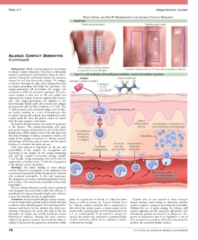
Plate 4-7                                                                                             Integumentary System

                                                           PATCH TESTING AND TYPE IV HYPERSENSITIVITY FOR ALLERGIC CONTACT DERMATITIS
                                                    Patch test
                                                         Patch test placement                        Positive patch test


       ALLERGIC CONTACT DERMATITIS
       (Continued)


         Pathogenesis: Much is known about the mechanism   Patch testing is the best method   Evaluation of patch tests at 72 hours shows papular erythema.
       of allergic contact dermatitis. This form of dermatitis   to assess for contact allergins.
       requires a sensitization and elicitation phase for devel-  Type IV (cell-mediated, delayed/hypersensitivity, contact dermatitis) reactions
       opment. During the sensitization phase, the patient is   Antigen           Ethylenediamine
       exposed for the first time to the antigen. The antigen   (allergen-carrier complex)
       is  absorbed  through  the  skin  and  is  phagocytosed  by
       an  antigen-presenting  cell  within  the  epidermis.  The
       antigen-presenting  cell  internalizes  the  antigen  and
       processes  it  within  its  lysosomal  apparatus.  The  pro-  Skin
       cessed  antigen  is  then  sent  to  the  cell  surface  and
       expressed on a human leukocyte antigen (HLA) mole-
       cule.  The  antigen-presenting  cell  migrates  to  the
       local  draining  lymph  node  and  presents  the  antigen
       in association with the HLA molecule to T cells. The
       T cells recognize each individual antigen and prolifer-              Antigen-presenting cell
       ate  locally,  resulting  in  a  clone  of  lymphocytes  that
       recognize that specific antigen; these lymphocytes then
       remain  ready  for  when  the  patient  comes  in  contact
       with the same antigen in the future.                                                     Edema,
         During the elicitation phase, the patient is reexposed                               inflammation,  Accumulation of lymphocytes
       to  the  antigen.  The  antigen-presenting  cells  again                                 fibrosis  and monocytes/macrophages
       process the antigen and present it to the newly cloned   T lymphocyte
       lymphocytes, which migrate back to the skin and cause   (previously  T
       the clinical findings of edema, spongiosis, vesicles, and   sensitized                     Fibroblast
       bullae. If the antigen is exposed in a chronic manner,   to antigen)
       the findings will be less acute in nature, and the typical
       findings of a chronic dermatitis are seen.                              Increased  Expression of
         This  entire  process  is  dependent  on  the  size  and   Cytokines   vascular  adhesion
       permeability  of  the  antigen,  the  recognition  and     Lymphokines  permeability  molecules
       processing  of  the  antigen  by  the  antigen-presenting   Blood vessel
       cell,  and  the  complex  interactions  among  multiple
       T and B cells. Antign-presenting cells and B cells are
       required for activation of the T cells and propagation
       of the allergic contact dermatitis.
         Histology:  The  initial  finding  in  acute  allergic
       contact dermatitis is spongiosis of the epidermis with
       an associated superficial and deep lymphocytic infiltrate                                          Margination and extra-
       with  scattered  eosinophils.  As  the  rash  progresses,                                          vasation of monocytes
       the spongiosis can worsen, and intraepidermal vesicles   Monocyte                                    and lymphocytes
       start to form. The vesicles may eventually coalesce into   Lymphocyte
       large bullae.
         Chronic allergic dermatitis usually shows acanthosis
       with spongiosis and eosinophils within the infiltrate. A
       superficial and deep perivascular lymphocytic infiltrate
       is seen. Excoriations can also be appreciated.
         Treatment: Acute localized allergic contact dermati-  plant.  As  a  good  rule  of  thumb,  if  a  plant  has  three   Patients  who  do  not  respond  to  these  measures
       tis can be treated with a potent topical steroid and strict   leaves, it could be poison ivy: “Leaves of three, let it   should  undergo  patch  testing  to  determine  whether
       avoidance of the offending agent. Oral sedating antihis-  be.”  Allergic  contact  dermatitis  that  is  widespread  or   another antigen is causing or provoking the dermatitis.
       tamines  work  better  for  the  pruritus  than  their  non-  that affects the eyelids, hands, or groin region can be   Without  the  use  of  patch  testing,  the  allergen  will
       sedating  counterparts  do.  Soaks  that  help  to  dry  the   treated with a tapering dose of oral corticosteroid over   remain  unknown  and  the  dermatitis  will  persist.  Not
       dermatitis  are  helpful  and  include  aluminum  acetate   a  2-  to  3-week  period.  If  the  steroid  is  tapered  too   infrequently, patients are found to be allergic to a fra-
       (Domeboro’s  solution).  Because  the  most  common   quickly, the patient may experience a poststeroid flare   grance or preservative that is an ingredient in one of
       culprit is the poison ivy plant, time should be taken to   of  their  dermatitis,  which  can  be  resistant  to  further   their personal care products. Once they stop using the
       explain to the patient the appearance and nature of this   corticosteroid therapy.  product, the dermatitis finally resolves.

       78                                                                                    THE NETTER COLLECTION OF MEDICAL ILLUSTRATIONS