
Page 93 - The Netter Collection of Medical Illustrations - Integumentary System_ Volume 4 ( PDFDrive )
P. 93
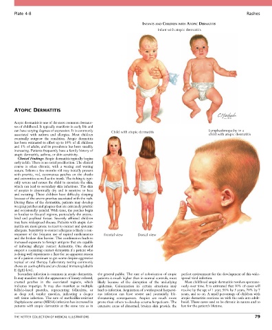
Plate 4-8                                                                                                            Rashes

                                                                         INFANTS AND CHILDREN WITH ATOPIC DERMATITIS
                                                                                Infant with atopic dermatitis


        ATOPIC DERMATITIS


        Atopic dermatitis is one of the most common dermato-
        ses of childhood. It typically manifests in early life and
        can have varying degrees of expression. It is commonly   Child with atopic dermatitis             Lymphadenopathy in a
        associated  with  asthma  and  allergies.  Most  children                                         child with atopic dermatitis
        eventually  outgrow  the  condition.  Atopic  dermatitis
        has been estimated to affect up to 10% of all children
        and 1% of adults, and its prevalence has been steadily
        increasing. Patients frequently have a family history of
        atopic dermatitis, asthma, or skin sensitivity.
          Clinical Findings: Atopic dermatitis typically begins
        early in life. There is no racial predilection. The clinical
        course  is  often  chronic,  with  a  waxing  and  waning
        nature. Infants a few months old may initially present
        with  pruritic,  red,  eczematous  patches  on  the  cheeks
        and extremities as well as the trunk. The itching is typi-
        cally severe and causes the child to excoriate the skin,
        which can lead to secondary skin infections. The skin
        of  atopics  is  abnormally  dry  and  is  sensitive  to  heat
        and  sweating.  These  children  have  difficulty  sleeping
        because of the severe pruritus associated with the rash.
        During  flares  of  the  dermatitis,  patients  may  develop
        weeping patches and plaques that are extremely pruritic
        and occasionally painful. With time, the patches begin
        to localize to flexural regions, particularly the antecu-
        bital  and  popliteal  fossae.  Severely  afflicted  children
        may have widespread disease. Patients with atopic der-
        matitis are more prone to react to contact and systemic
        allergens. Sensitivity to contact allergens is likely a con-
        sequence  of  the  frequent  use  of  topical  medicaments   Frontal view  Dorsal view
        and the broken skin barrier. This combination leads to
        increased exposure to foreign antigens that are capable
        of  inducing  allergic  contact  dermatitis.  One  should
        suspect a coexisting contact dermatitis if a patient who
        is doing well experiences a flare for no apparent reason
        or if a patient continues to get worse despite aggressive
        topical or oral therapy. Laboratory testing commonly
        shows an eosinophilia and an elevated immunoglobulin
        E (IgE) level.
          Secondary infection is common in atopic dermatitis.   the general public. The rate of colonization of atopic   perfect environment for the development of this wide-
        It may manifest with the appearance of honey-colored,   patients is much higher than in normal controls, most   spread viral infection.
        crusted  patches  in  the  excoriated  regions,  which     likely  because  of  the  disruption  of  the  underlying     Most childhood atopic dermatitis resolves spontane-
        indicates  impetigo.  It  may  also  manifest  as  multiple   epidermis.  Colonization  in  certain  situations  may     ously over time. It is estimated that 10% of cases will
        follicle-based  pustules,  representing  folliculitis,  or     lead to infection. Acquisition of a widespread herpesvi-  resolve by the age of 1 year, 50% by 5 years, 70% by 7
        as  deep  red,  tender  macules,  indicating  a  deeper     rus  infection  can  have  severe  and  potentially  life-  years, and so on. A small percentage of children with
        soft  tissue  infection.  The  rate  of  methicillin-resistant   threatening  consequences.  Atopics  are  much  more   atopic dermatitis continue on with the rash into adult-
        Staphylococcus aureus (MRSA) infection has increased in   prone than others to develop eczema herpeticum. The   hood. These cases tend to be chronic in nature and to
        patients  with  atopic  dermatitis  at  the  same  rate  as  in   extensive  areas  of  abnormal,  broken  skin  provide  the   last for the patient’s lifetime.


        THE NETTER COLLECTION OF MEDICAL ILLUSTRATIONS                                                                           79