
Page 94 - The Netter Collection of Medical Illustrations - Integumentary System_ Volume 4 ( PDFDrive )
P. 94
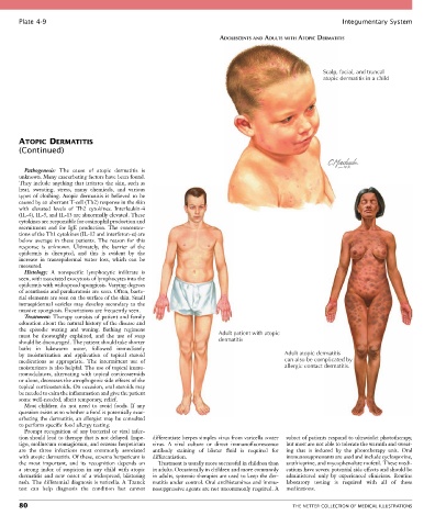
Plate 4-9                                                                                             Integumentary System

                                                                      ADOLESCENTS AND ADULTS WITH ATOPIC DERMATITIS


                                                                                                       Scalp, facial, and truncal
                                                                                                       atopic dermatitis in a child


       ATOPIC DERMATITIS
       (Continued)


         Pathogenesis:  The  cause  of  atopic  dermatitis  is
       unknown. Many exacerbating factors have been found.
       They include anything that irritates the skin, such as
       heat,  sweating,  stress,  many  chemicals,  and  various
       types  of  clothing.  Atopic  dermatitis  is  believed  to  be
       caused by an aberrant T-cell (Th2) response in the skin
       with  elevated  levels  of  Th2  cytokines.  Interleukin-4
       (IL-4), IL-5, and IL-13 are abnormally elevated. These
       cytokines are responsible for eosinophil production and
       recruitment  and  for  IgE  production.  The  concentra-
       tions of the Th1 cytokines (IL-12 and interferon-α) are
       below  average  in  these  patients.  The  reason  for  this
       response  is  unknown.  Ultimately,  the  barrier  of  the
       epidermis  is  disrupted,  and  this  is  evident  by  the
       increase  in  transepidermal  water  loss,  which  can  be
       measured.
         Histology:  A  nonspecific  lymphocytic  infiltrate  is
       seen, with associated exocytosis of lymphocytes into the
       epidermis with widespread spongiosis. Varying degrees
       of acanthosis and parakeratosis are seen. Often, bacte-
       rial elements are seen on the surface of the skin. Small
       intraepidermal  vesicles  may  develop  secondary  to  the
       massive spongiosis. Excoriations are frequently seen.
         Treatment:  Therapy  consists  of  patient  and  family
       education about the natural history of the disease and
       the  episodic  waxing  and  waning.  Bathing  regimens
       must  be  thoroughly  explained,  and  the  use  of  soap      Adult patient with atopic
       should be discouraged. The patient should take shorter         dermatitis
       baths  in  lukewarm  water,  followed  immediately
       by  moisturization  and  application  of  topical  steroid                         Adult atopic dermatitis
       medications  as  appropriate.  The  intermittent  use  of                          can also be complicated by
       moisturizers is also helpful. The use of topical immu-                             allergic contact dermatitis.
       nomodulators, alternating with topical corticosteroids
       or alone, decreases the atrophogenic side effects of the
       topical corticosteroids. On occasion, oral steroids may
       be needed to calm the inflammation and give the patient
       some well-needed, albeit temporary, relief.
         Most  children  do  not  need  to  avoid  foods.  If  any
       question exists as to whether a food is potentially exac-
       erbating the dermatitis, an allergist may be consulted
       to perform specific food allergy testing.
         Prompt  recognition  of  any  bacterial  or  viral  infec-
       tion should lead to therapy that is not delayed. Impe-  differentiate herpes simplex virus from varicella zoster   subset of patients respond to ultraviolet phototherapy,
       tigo, molluscum contagiosum, and eczema herpeticum   virus.  A  viral  culture  or  direct  immunofluorescence   but most are not able to tolerate the warmth and sweat-
       are  the  three  infections  most  commonly  associated   antibody  staining  of  blister  fluid  is  required  for   ing  that  is  induced  by  the  phototherapy  unit.  Oral
       with atopic dermatitis. Of these, eczema herpeticum is   differentiation.           immunosuppressants are used and include cyclosporine,
       the  most  important,  and  its  recognition  depends  on     Treatment is usually more successful in children than   azathioprine, and mycophenolate mofetil. These medi-
       a  strong  index  of  suspicion  in  any  child  with  atopic   in adults. Occasionally in children and more commonly   cations have severe potential side effects and should be
       dermatitis  and  new  onset  of  a  widespread,  blistering   in adults, systemic therapies are used to keep the der-  administered  only  by  experienced  clinicians.  Routine
       rash. The differential diagnosis is varicella. A Tzanck   matitis under control. Oral antihistamines and immu-  laboratory  testing  is  required  with  all  of  these
       test  can  help  diagnosis  the  condition  but  cannot   nosuppressive agents are not uncommonly required. A   medications.

       80                                                                                    THE NETTER COLLECTION OF MEDICAL ILLUSTRATIONS