
Page 376 - Cardiac Nursing
P. 376
1
1
2:1
/30
/09
/09
M
M
Pa
2:1
6 A
6 A
/30
87.
q
q
3-3
3-3
87.
6
6
6
q
xd
xd
ara
ara
a
p
t
t
c.
c.
c.
a
In
In
p
g
e 3
e 3
Pa
g
g
A
A
p
52
52
A
16_
16_
0-c
33
33
p
0-c
LWB
LWBK340-c16_ p pp333-387.qxd 6/30/09 12:16 AM Page 352 Aptara Inc.
LWB
K34
K34
352 P A R T III / Assessment of Heart Disease
Atrial Tachycardia (AT) In addition to the potential hemodynamic instability resulting
AT is a rapid atrial rhythm at a rate of 100 to 250 beats per from the rapid ventricular rate, AT, and other SVTs that result in
minute that arises from a single site within the right or left atrium. rapid ventricular rates for long periods of time can cause tachycardia-
24
This rhythm may be due to rapid firing of an ectopic atrial focus mediated cardiomyopathy. Chronic tachycardia produces complex
(automaticity), an atrial microreentry circuit that allows an im- structural changes and “remodeling” of both the atria and the ven-
pulse to travel rapidly and repeatedly around a pathway in the tricles. Left ventricular dilation can lead to dilated cardiomyopathy
atria, or to afterdepolarizations resulting in a triggered AT. 20–23 and systolic dysfunction. For this reason, chronic tachycardia needs
The term paroxysmal atrial tachycardia is used to describe AT that to be treated to avoid development of cardiomyopathy.
begins and ends suddenly and can occur in short bursts of several Treatment of AT is directed toward eliminating the cause,
beats or be sustained for longer periods of time. Incessant AT is decreasing the ventricular rate, and ultimately preventing recur-
less common and lasts for more than half a day, sometimes being rences of tachycardia. Sedation alone may terminate the rhythm
23
present more than 90% of the time. AT has been associated with or slow the rate. Vagal stimulation, either through CSM or Val-
caffeine, tobacco, alcohol, mitral valve disease, rheumatic heart salva maneuver, or adenosine may terminate some episodes of
disease, chronic obstructive pulmonary disease, acute MI, theo- AT. -Blockers, verapamil, and diltiazem increase block at the
phylline administration, hypokalemia, and digitalis toxicity. AV node and may slow ventricular response or sometimes ter-
If the atrial rate is very rapid, the AV node begins to block minate the tachycardia. Digitalis slows ventricular rate by in-
some of the impulses attempting to travel through it to protect the creasing block at the AV node, but it can also be the cause of AT
ventricles from excessively rapid rates. In normal, healthy hearts, with block and should be discontinued if that is the case. If the
the AV node can usually conduct each atrial impulse up to rates patient cannot tolerate -blockers or calcium channel blocker,
of 180 beats per minute or more. In patients with cardiac disease IV amiodarone can control ventricular rate and may convert the
or in those who take drugs that slow AV conduction, the AV node rhythm to sinus. Other antiarrhythmics that might be effective
25
cannot conduct each impulse, and AT with block occurs. The include flecainide, propafenone, procainamide, or sotalol ;
presence of AT with block should arouse suspicion of digitalis tox- however, all carry a risk of proarrhythmia, which is greater than
icity, which must be ruled out. the risk with amiodarone. If the ventricular rate is so fast that
The ECG characteristics of AT include the following: hemodynamic instability occurs, then cardioversion can be at-
tempted. Cardioversion is usually not effective in managing AT
Rate: Atrial rate is 100 to 250 beats per minute (quite often in the
that is due to enhanced automaticity. Radiofrequency catheter
range of 140 to 180 beats per minute). The ventricular rate de-
ablation of the ectopic focus or reentry circuit is now a primary
pends on the amount of block at the AV node and may be the
therapy for AT, with success rates varying from 52% to 98% de-
same as the atrial rate or slower.
pending on the site of AT; and a recurrence rate of about
Rhythm: Regular unless there is variable block at the AV node 21,25,26
8%.
P waves: Differ in configuration from sinus P waves because they
are ectopic. Precede each QRS complex and usually appear in Atrial Flutter
the second half of the tachycardia cycle (R-R interval) but may
Atrial flutter is an organized atrial rhythm in which the atria are
be hidden in the preceding T wave. When block is present,
depolarized at rates of 250 to 440 times per minute. Classic or
more than one P wave appears before each QRS complex.
typical atrial flutter (type I) is due to a fixed reentry circuit in the
PR interval: Usually in the normal range but often difficult to
right atrium around which the impulse circulates in a counter-
measure because of hidden P waves.
clockwise direction, resulting in negative flutter waves in leads II
QRS complex: Usually normal but may be wide if aberrant con-
and III and an atrial rate between 250 and 350 beats per minute
duction is present 25,27,28
(most commonly 300 beats per minute). Occasionally, the
Conduction: Usually normal through the AV node and into the
impulse reverses direction and circulates in a clockwise direction,
ventricles. In AT with block, some atrial impulses do not con-
resulting in positive flutter waves in leads II and III, and is called
duct into the ventricles. Aberrant ventricular conduction may 27,28
“atypical” or “reverse typical” flutter. Atrial flutter can also re-
occur if atrial impulses are conducted into the ventricles while
sult from reentry around surgically created scars within the atria
the bundle branches are still partially refractory.
and is still considered to be type I flutter. Less is known about type
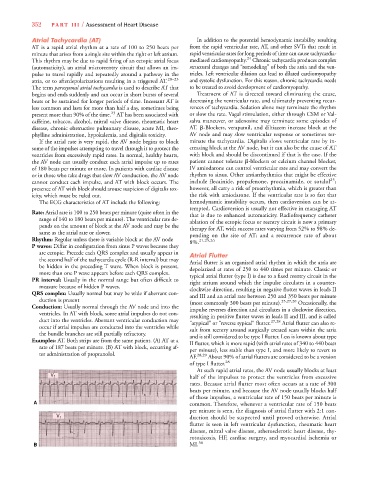
Examples: AT. Both strips are from the same patient. (A) AT at a
II flutter, which is more rapid (with atrial rates of 340 to 440 beats
rate of 187 beats per minute. (B) AT with block, occurring af-
per minute), less stable than type I, and more likely to revert to
ter administration of propranolol. 28,29
AF. About 90% of atrial flutters are considered to be a version
of type I flutter. 28
At such rapid atrial rates, the AV node usually blocks at least
half of the impulses to protect the ventricles from excessive
V 1
rates. Because atrial flutter most often occurs at a rate of 300
beats per minute, and because the AV node usually blocks half
of those impulses, a ventricular rate of 150 beats per minute is
A common. Therefore, whenever a ventricular rate of 150 beats
per minute is seen, the diagnosis of atrial flutter with 2:1 con-
duction should be suspected until proved otherwise. Atrial
V 1
flutter is seen in left ventricular dysfunction, rheumatic heart
disease, mitral valve disease, atherosclerotic heart disease, thy-
rotoxicosis, HF, cardiac surgery, and myocardial ischemia or
B MI. 30