
Page 377 - Cardiac Nursing
P. 377
1
1
2:1
/09
/09
1
M
M
Pa
2:1
6 A
6 A
/09
q
q
q
3-3
87.
87.
6
/30
/30
xd
xd
6
t
ara
ara
p
p
t
In
c.
c.
a
a
In
p
g
g
e 3
Pa
Pa
g
53
A
A
e 3
53
53
3-3
0-c
K34
16_
33
33
LWB K34 0-c 16_ p p pp333-387.qxd 6/30/09 12:16 AM Page 353 Aptara Inc.
LWB
LWBK340-c16_
C HAPTER 1 6 / Arrhythmias and Conduction Disturbances 353
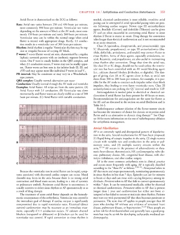
Atrial flutter is characterized on the ECG as follows: needed, electrical cardioversion is most reliable; overdrive atrial
pacing can be attempted if atrial epicardial pacing wires are pres-
Rate: Atrial rate varies between 250 and 450 beats per minute,
ent following cardiac surgery. Newer type III antiarrhythmic
most commonly 300 beats per minute. Ventricular rate varies
agents, ibutilide (Corvert) and dofetilide (Tikosyn) can be given
depending on the amount of block at the AV node, most com-
IV and are often successful in converting atrial flutter to sinus
monly 150 beats per minute and rarely 300 beats per minute.
rhythm if flutter is recent in onset. Drug therapy for conversion
Ventricular rates can be within the normal range when atrial
takes longer than electrical cardioversion and is not recommended
flutter is treated with appropriate drugs. Rarely, 1:1 conduc-
in an acute situation.
tion results in a ventricular rate of 300 beats per minute.
Class IA (quinidine, disopyramide, and procainamide), type
Rhythm: Atrial rhythm is regular. Ventricular rhythm may be reg-
IC (flecainide, propafenone), or type III antiarrhythmics (ibu-
ular or irregular because of varying AV block.
tilide, dofetilide, amiodarone, and sotalol) may convert flutter to
P waves: F waves (flutter waves) are seen, characterized by a regular,
sinus rhythm. Some of these agents, especially amiodarone, so-
biphasic sawtooth pattern with no isoelectric segment between
talol, flecainide, and propafenone, are also useful in maintaining
waves. One F wave is usually hidden in the QRS complex, and
sinus rhythm after conversion. Drugs that slow the atrial rate,
when 2:1 conduction occurs, F waves may not be readily appar-
like class IA or IC drugs, should not be used unless the ventric-
ent. Flutter waves are best seen in the inferior leads (II, III, and
ular rate has been controlled with an AV nodal blocking agent
aVF) and may appear more like individual P waves in lead V 1 .
(i.e., calcium channel blocker, -blocker, or digitalis). The dan-
PR interval: May be consistent or may vary in a Wenckebach-
ger of giving class IA or IC agents alone is that, as atrial rate
type pattern.
slows from 300 to 200 beats per minute, for example, it is pos-
QRS complex: Usually normal; aberration can occur
sible for the AV node to conduct each impulse rather than block
Conduction: Usually normal through the AV node and ventricles
impulses, thus leading to even faster ventricular rates. Class III
Examples: Atrial flutter. All strips are from the same patient. (A)
antiarrhythmics can prolong the QT interval and result in TdP.
Atrial flutter with 2:1 conduction. (B) Ventricular rate slows
Anticoagulation is needed prior to electrical or chemical car-
momentarily, and flutter waves are clearly visible at a rate of 300
dioversion if atrial flutter has been present for 48 hours or more.
beats per minute. (C) Atrial flutter with variable conduction.
Recommendations for anticoagulation are the same for flutter as
for AF, and are discussed in the section on atrial fibrillation and in
V 1 Table 16-3.
Radiofrequency catheter ablation of the flutter reentry circuit
has become the treatment of choice for chronic or recurrent atrial
flutter and is an alternative to chronic drug therapy. 31 See Chap-
ter 18 for more information on the use of radiofrequency ablation
A
for arrhythmia management.
V 1 Atrial Fibrillation
AF is an extremely rapid and disorganized pattern of depolariza-
tion in the atria. Several mechanisms for AF have been proposed:
(1) Rapid firing of ectopic impulses in the atria, (2) single reentry
B circuit with variable rate and conduction in the atria or pul-
monary veins, and (3) multiple reentry circuits within the
V 1 atria. 32–34 AF occurs in the presence of atherosclerotic or rheu-
matic heart disease, thyrotoxicosis, HF, cardiomyopathy, valve dis-
ease, pulmonary disease, MI, congenital heart disease, with elec-
trolyte imbalances, and after cardiac surgery.
AF is the most common arrhythmia seen in clinical practice
C
and occurs more frequently with aging. AF is typically classified
according to the “three Ps” as follows. 5,32–35 : Paroxysmal refers to
Because the ventricular rate in atrial flutter can be rapid, symp- AF that starts and stops spontaneously, terminating spontaneously
toms associated with decreased cardiac output can occur. Mural in sinus rhythm in less than 7 days. Episodes can last for minutes
thrombi may form in the atria because there is no strong atrial to hours or days and can recur with varying frequency among in-
contraction and blood stasis occurs, leading to a risk of systemic dividuals. Persistent refers to AF that fails to convert spontaneously
or pulmonary emboli. Persistent atrial flutter is uncommon; it within 7 days. It can be terminated medically, either by electrical
usually converts to either sinus rhythm or AF spontaneously or as or chemical cardioversion. Permanent refers to AF that is present
a result of drug therapy. longer than 1 year and cardioversion has either not been at-
The treatment of acute atrial flutter depends on the hemody- tempted or has failed to restore or maintain sinus rhythm. Patients
namic consequences of the arrhythmia. Ventricular rate control is may start out with paroxysmal AF that then becomes persistent or
the immediate goal of therapy if cardiac output is significantly permanent. The term lone AF applies to people younger than 60
compromised due to rapid ventricular rates. Electrical (direct years who develop AF without any evidence of structural heart
current) cardioversion may be necessary as an immediate treat- disease, pulmonary disease, or hypertension. 34,35 These people are
ment, especially if 1:1 conduction occurs. IV calcium channel at low risk for thromboembolism and generally have a good prog-
blockers (verapamil or diltiazem) or -blockers can be used for nosis but may be at risk for developing tachycardia-mediated car-
ventricular rate control. If rapid conversion to sinus rhythm is diomyopathy.