
Page 378 - Cardiac Nursing
P. 378
1
2:1
2:1
/09
1
1
M
Pa
Pa
6 A
6 A
M
q
xd
xd
87.
q
q
/30
/09
/09
6
6
/30
t
ara
ara
p
p
t
In
c.
c.
a
a
In
g
e 3
e 3
Pa
g
g
A
A
p
54
54
54
87.
16_
0-c
33
33
LWB
LWB K34 0-c 16_ p p pp333-387.qxd 6/30/09 12:16 AM Page 354 Aptara Inc.
K34
LWBK340-c16_
3-3
3-3
354 P A R T III / Assessment of Heart Disease
AF is characterized on the ECG as follows: diac output. Another possible complication is mural thrombus
formation in the atria due to stasis of blood, leading to pul-
Rate: Atrial rate is 400 to 600 beats per minute or faster. Ventric-
monary or systemic embolization if clots dislodge spontaneously
ular rate varies depending on the amount of block at the AV
or with conversion to sinus rhythm. Any tachyarrhythmia that is
node. In new-onset AF, the ventricular response is usually rapid,
sustained for long periods of time can lead to tachycardia-medi-
110 to 160 beats per minute; in treated AF, the ventricular rate
ated (or tachycardia induced) cardiomyopathy with ventricular
is controlled in the normal range of 60 to 100 beats per minute.
dilation and reduced LV function. The mechanism of this car-
Rhythm: Irregular. One of the distinguishing features of AF is the
diomyopathy is not well understood, but improvement in left
marked irregularity of the ventricular response because of
ventricular function is seen with ventricular rate control or
concealed conduction in the AV junction. If the ventricular re- 24
restoration of sinus rhythm.
sponse is ever regular in the presence of AF, AV dissociation
Treatment of AF is directed toward eliminating the cause, con-
should be suspected.
trolling ventricular rate, restoring and maintaining sinus rhythm
P waves: Not present. Atrial activity is chaotic, with no formed
if possible, and preventing thromboembolism. Emergent or ur-
atrial impulses visible. Irregular F waves are often seen and vary
gent electrical cardioversion may be necessary if the patient is he-
in size from coarse to very fine.
modynamically unstable because of a rapid ventricular rate. Car-
PR interval: Not measurable because there are no P waves
dioversion is more likely to be successful if AF has been present for
QRS complex: Usually normal; aberration is common, especially
less than 24 hours.
at faster ventricular rates
Two management strategies are available for patients in AF:
Conduction: Intra-atrial conduction is disorganized and irreg-
rate control or rhythm control. Rate control means that ventricu-
ular. Most of the atrial impulses are blocked in the AV junc-
lar rate is controlled with drug therapy with no intent to restore
tion; those impulses that are conducted through the AV
sinus rhythm. Rhythm control means that treatment is aimed at
junction are usually conducted normally through the ven-
restoring and maintaining sinus rhythm. Traditionally it has been
tricles. If an atrial impulse reaches the bundle-branch sys-
thought that restoration of sinus rhythm would result in better
tem during its refractory period, aberrant intraventricular
outcomes because atrial kick would be restored, hemodynamics
conduction can occur.
would improve, the incidence of stroke and of bleeding would be
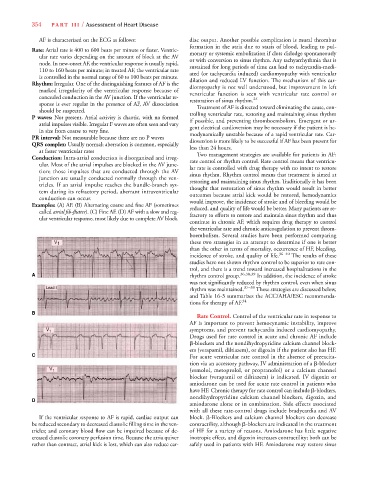
Examples: (A) AF. (B) Alternating coarse and fine AF (sometimes
reduced, and quality of life would be better. Many patients are re-
called atrial fib-flutter). (C) Fine AF. (D) AF with a slow and reg-r r
fractory to efforts to restore and maintain sinus rhythm and thus
ular ventricular response, most likely due to complete AV block.
continue in chronic AF, which requires drug therapy to control
the ventricular rate and chronic anticoagulation to prevent throm-
boembolism. Several studies have been performed comparing
V 1 these two strategies in an attempt to determine if one is better
than the other in terms of mortality, occurrence of HF, bleeding,
incidence of stroke, and quality of life. 36–40 The results of these
studies have not shown rhythm control to be superior to rate con-
trol, and there is a trend toward increased hospitalizations in the
A A rhythm control group. 36,38,39 In addition, the incidence of stroke
was not significantly reduced by rhythm control, even when sinus
rhythm was maintained. 37–39 These strategies are discussed below,
and Table 16-3 summarizes the ACC/AHA/ESC recommenda-
tions for therapy of AF. 34
B
Rate Control. Control of the ventricular rate in response to
AF is important to prevent hemocynamic instability, improve
V 1 symptoms, and prevent tachycardia induced cardiomyopathy.
Drugs used for rate control in acute and chronic AF include
-blockers and the nondihydropyridine calcium channel block-
ers (verapamil, diltiazem), or digoxin if the patient also has HF.
C C For acute ventricular rate control in the absence of preexcita-
tion via an accessory pathway, IV administration of a -blocker
V 1 (esmolol, metoprolol, or propranolol) or a calcium channel
blocker (verapamil or diltiazem) is indicated. IV digoxin or
amiodarone can be used for acute rate control in patients who
have HF. Chronic therapy for rate control can include -blockers,
nondihydropyridine calcium channel blockers, digoxin, and
D D
amiodarone alone or in combination. Side effects associated
with all these rate-control drugs include bradycardia and AV
If the ventricular response to AF is rapid, cardiac output can block. -Blockers and calcium channel blockers can decrease
be reduced secondary to decreased diastolic filling time in the ven- contractility, although -blockers are indicated in the treatment
tricles; and coronary blood flow can be impaired because of de- of HF for a variety of reasons. Amiodarone has little negative
creased diastolic coronary perfusion time. Because the atria quiver inotropic effect, and digoxin increases contractility; both can be
rather than contract, atrial kick is lost, which can also reduce car- safely used in patients with HF. Amiodarone may restore sinus