
Page 382 - Cardiac Nursing
P. 382
1
1
1
/09
/09
/09
6 A
M
M
2:1
2:1
6 A
/30
87.
q
q
3-3
3-3
87.
6
6
/30
q
xd
xd
Pa
t
ara
ara
p
p
t
In
c.
c.
a
a
In
p
g
g
e 3
Pa
Pa
g
58
A
A
e 3
58
58
33
p
LWB
33
0-c
16_
p
K34
LWBK340-c16_
LWB K34 0-c 16_ pp333-387.qxd 6/30/09 12:16 AM Page 358 Aptara Inc.
358 P A R T III / Assessment of Heart Disease
When the sinus rhythm is undisturbed by PVCs, the atria de- The treatment of accelerated ventricular rhythm depends on
polarize normally. its cause and how well it is tolerated by the patient. This arrhyth-
Examples: (A) Normal sinus rhythm with a PVC. (B) Sinus mia alone is usually not harmful because the ventricular rate is
rhythm with multifocal PVC. (C) Paired PVC. (D) R-on-T within normal limits and usually adequate to maintain cardiac
PVC, resulting in short runs of VT. output. If the patient is symptomatic because of the loss of atrial
kick during long episodes of AV dissociation, atropine can be used
The significance of PVCs depends on the clinical setting in
to increase the rate of the SA node and overdrive the ventricular
which they occur. Many people have chronic PVCs that do not
rhythm. Suppressive therapy is rarely used because abolishing the
need to be treated, and most of these people are asymptomatic.
ventricular rhythm may leave an even less desirable heart rate.
There is no evidence that suppression of PVCs reduces mortality,
Usually, accelerated ventricular rhythm is transient and benign
especially in patients with no structural heart disease. If PVCs
and does not require treatment.
cause bothersome palpitations, patients should be told to avoid
caffeine, tobacco, other stimulants, and try stress reduction tech- Ventricular Tachycardia
niques. Low-dose -blockers may reduce PVC frequency and the VT is a rapid ventricular rhythm most likely due to reentry in the
perception of palpitations and can be used for symptom relief. In ventricles, although automaticity of an ectopic focus and afterde-
the setting of an acute MI or myocardial ischemia, PVCs may be polarizations may also be mechanisms of VT. 5,22 VT can be clas-
precursors of more dangerous ventricular arrhythmias, especially sified according to (1) duration—nonsustained (lasts 30 sec-
when they occur near the apex of the T wave (R-on-T PVC). Pro- onds), sustained (lasts 30 seconds), incessant (VT present most
phylactic treatment of asymptomatic nonsustained ventricular ar- of the time); (2) morphology (ECG appearance of QRS com-
rhythmias is not recommended. 49 plexes)—monomorphic (QRS complexes have the same shape dur-
ing tachycardia), polymorphic (QRS complexes vary randomly in
Accelerated Idioventricular Rhythm shape), bidirectional (alternating upright and negative QRS com-
Accelerated idioventricular rhythm occurs when an ectopic focus plexes during tachycardia). The terms salvos and bursts are often
in the ventricles fires at a rate of 50 to 100 beats per minute. Ac- used to describe short runs of VT (i.e., 5 to 10 or more beats in a
celerated idioventricular rhythm commonly occurs in the pres- row). See section titled “Complex Arrhythmias and Conduction
ence of inferior MI and during reperfusion with thrombolytic Disturbances” later in this chapter for more information on
therapy, when the rate of the SA node slows below the rate of the monomorphic and PVT.
latent ventricular pacemaker. (See section titled “Complex Ar- The most common cause of VT is CHD, including acute is-
rhythmias and Conduction Disturbances” for a discussion of AV chemia and MI, prior MI, and chronic coronary disease. The next
dissociation.) The ECG characteristics of accelerated ventricular most common cause is cardiomyopathy, both dilated and hyper-
rhythm include the following: trophic. Other causes include valvular heart disease, congenital
heart disease, arrhythmogenic right ventricular dysplasia, inher-
Rate: 50 to 100 beats per minute
Rhythm: Usually regular ited ion channel abnormalities, cardiac surgery, and the proar-
22,48,50,51
P waves: May be seen but are dissociated from the QRS. If retro- rhythmic effects of many drugs. VT that occurs in the
grade conduction from the ventricle to the atria occurs, P presence of left ventricular dysfunction and reduced ejection frac-
waves follow the QRS complex. tion is associated with a higher incidence of adverse cardiac events,
PR interval: Not present including an increased risk of SCD.
QRS complex: Wide and bizarre Idiopathic VT is VT that occurs in patients with no known
22,48,52
Conduction: If sinus rhythm is the basic rhythm, atrial conduc- structural heart disease. This type of VT is discussed in
tion is normal. Impulses originating in the ventricles conduct more detail later in the section titled “Complex Arrhythmias and
through the ventricular myocardium by cell-to-cell conduc- Conduction Disturbances”.
tion, resulting in the wide QRS complex. ECG characteristics of monomorphic VT include the follow-
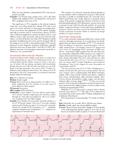
Example: Sinus rhythm with accelerated ventricular rhythm at a ing:
rate of 70 beats per minute. Note sinus P waves that continue Rate: Ventricular rate is usually 100 to 220 beats per minute
uninterrupted during the period of accelerated ventricular Rhythm: Usually regular but may be slightly irregular
rhythm (an example of AV dissociation). (N arrhythmia P waves: Often dissociated from QRS complexes. If sinus rhythm
computer’s interpretation of normal beat, V computer’s in- is the underlying basic rhythm, regular P waves may be seen
terpretation of ventricular beat.) but are not related to QRS complexes. P waves are often buried
Example of accelerated ventricular rhythm