
Page 384 - Cardiac Nursing
P. 384
1
1
2:1
/30
/09
/09
M
M
Pa
2:1
6 A
6 A
/30
87.
q
q
3-3
3-3
87.
6
6
6
q
xd
xd
ara
ara
a
p
t
t
c.
c.
c.
a
In
In
p
g
e 3
e 3
Pa
g
g
A
A
p
60
60
A
LWBK340-c16_
33
K34
16_
0-c
LWB
LWB K34 0-c 16_ p p pp333-387.qxd 6/30/09 12:16 AM Page 360 Aptara Inc.
33
360 P A R T III / Assessment of Heart Disease
VF requires immediate defibrillation. Synchronized cardiover- CPR must be initiated immediately if the patient is to survive.
sion is not possible because there are no formed QRS complexes IV epinephrine and atropine may be given in an effort to stimu-
on which to synchronize the shock. CPR must be performed if a late a rhythm; vasopressin can be used instead of epinephrine as a
defibrillator is not immediately available. The American Heart As- vasopressor. Asystole has a very poor prognosis despite the best re-
sociation guidelines for VF and pulseless VT call for CPR until a suscitation efforts because it usually represents extensive myocar-
defibrillator is available, then immediate defibrillation using and dial ischemia or severe underlying metabolic problems. See Chap-
automatic external defibrillator; or manual defibrillation with 360 J ter 27 for the ACLS algorithm for treatment of asystole.
if using a monophasic defibrillator or device-specific energy rec-
ommendation (200 J if this is not known) if using a biphasic de- Conduction Abnormalities
fibrillator. 53 Immediate CPR for 2 minutes is recommended fol-
lowing the first shock. See Chapter 27 for more information on The term AV block is used to describe arrhythmias in which there
management of cardiac arrest. is delayed or failed conduction of supraventricular impulses into
Amiodarone is the drug recommended for antiarrhythmic the ventricles. AV blocks have been classified according to location
therapy in VF following defibrillation. Lidocaine is an alternative of the block and severity of the conduction abnormality. The fol-
(but not the preferred drug) according to the ACLS manual and lowing classification of AV blocks is discussed in this section:
is still used clinically in many hospitals. Drugs have not been
shown to improve survival in patients with recurrent hemody- First-degree AV block
namically unstable ventricular arrhythmias; even amiodarone, Second-degree AV block
which is the most effective antiarrhythmic, is inferior to ICD in Type I
reducing the incidence of SCD. However, amiodarone and - Type II
blockers, often in combination, are used in patients with recurrent 2:1 conduction (can be type I or type II)
ventricular arrhythmias who are not eligible for ICD implantation High-grade AV block (or advanced AV block)
or in those who have an ICD but have recurrent ventricular ar- Third-degree AV block
rhythmias that cause frequent ICD shocks. Sotalol is also effective AV block can be caused by disease processes that either inter-
in suppressing ventricular arrhythmias in many patients. rupt the blood supply to structures in the conduction system or
otherwise interfere with the function of these structures, or by
Ventricular Asystole drugs that slow conduction through the AV node. It can also oc-
Ventricular asystole is the absence of any ventricular rhythm; there cur in normal hearts and be a result of normal physiologic varia-
is no QRS complex, no pulse, and no cardiac output. The term tions (e.g., vagal tone) that affect conduction through the AV
“ventricular standstill” is sometimes used when atrial activity is node, or in athletes or people who exercise regularly; it can occur
still present but no ventricular activity occurs. Both situations are during sleep when sympathetic tone is reduced or vagal tone is en-
fatal unless treated immediately. Ventricular asystole has the fol- hanced. One of the main functions of the AV node is to block
lowing characteristics: rapid atrial impulses to prevent dangerously fast ventricular rates in
Rate: None response to rapid atrial rhythms such as rapid AT, atrial flutter, or
Rhythm: None AF. In this case, the block is physiologic and must not be confused
P waves: May be present if the SA node is functioning with pathologic block due to abnormal AV node function. For ex-
PR interval: None ample, a sinus rate of 80 should be conducted through a normally
QRS complex: None functioning AV conduction system in a 1:1 fashion, so, if any of
Conduction: Atrial conduction may be normal if the SA node is those sinus impulses are blocked, that is abnormal AV node func-
functioning. There is no conduction into the ventricles. tion and the term block appropriately applies. However, atrial flut-
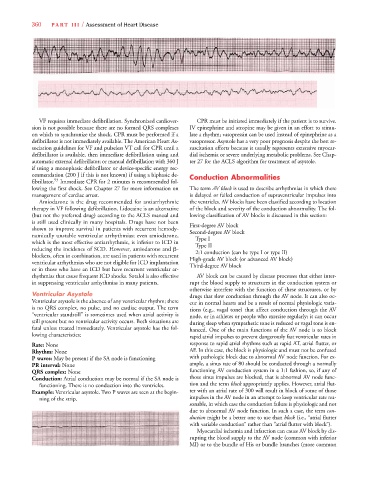
Example: Ventricular asystole. Two P waves are seen at the begin- ter with an atrial rate of 300 will result in block of some of those
ning of the strip. impulses in the AV node in an attempt to keep ventricular rate rea-
sonable, in which case the conduction failure is physiologic and not
due to abnormal AV node function. In such a case, the term con-
duction might be a better one to use than block (i.e., “atrial flutter
with variable conduction” rather than “atrial flutter with block”).
Myocardial ischemia and infarction can cause AV block by dis-
rupting the blood supply to the AV node (common with inferior
MI) or to the bundle of His or bundle branches (more common