
Page 387 - Cardiac Nursing
P. 387
1
1
2:1
1
ara
a
/09
2:1
p
a
In
p
6 A
33
33
ara
6
6
xd
/30
/09
/09
/30
xd
87.
3-3
3-3
87.
q
q
q
In
63
63
63
A
A
g
g
g
e 3
e 3
p
K34
0-c
LWBK340-c16_
LWB K34 0-c 16_ pp333-387.qxd 6/30/09 12:16 AM Page 363 Aptara Inc.
LWB
p
p
t
16_
t
Pa
6 A
M
M
c.
Pa
Pa
c.
C HAPTER 1 6 / Arrhythmias and Conduction Disturbances 363
type I is normal on conducted beats because the blocked P Conduction: Normal through the atria. Two or more con-
wave allows enough time for the AV node to recover so that secutive atrial impulses fail to conduct to the ventricles.
it is able to conduct every other P wave with a normal PR Conduction through the ventricles is normal if block oc-
interval. If there are any periods of typical Wenckebach con- curs in the AV node and slow if block occurs in the bun-
duction with progressive lengthening of the PR interval on dle branches.
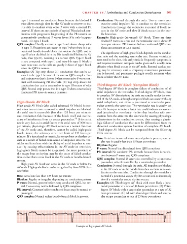
consecutively conducted P waves (even if it only happens Example: High-grade (advanced) AV block. There are three
once), it is type I block. blocked P waves in a row and the ventricular rate is about 25
QRS complex: Usually narrow in type I and almost always wide beats per minute. PR intervals before conducted QRS com-
in type II. Exceptions can occur in type I when there is a co- plexes are constant at 0.32 second.
incidental bundle-branch block that widens the QRS, and in The significance of high-grade block depends on the conduc-
type II when the block is in the His bundle (still below the AV tion ratio and the resulting ventricular rate. Because ventricular
node, thus type II), resulting in a narrow QRS. Type II block rates tend to be slow, this arrhythmia is frequently symptomatic
is rare compared with type I, and intra-His type II block is and requires treatment. Atropine can be given and is usually more
even more rare, so the odds are greatly in favor of type I block effective when block occurs in the AV node. External cardiac pac-
when the QRS is narrow. ing may be necessary until a temporary transvenous pacemaker
Examples: (A) Top strip shows 2:1 conduction that can be as- can be inserted, and permanent pacing is usually necessary when
sumed to be type I because of the narrow QRS complex. Sec- block is below the AV node.
ond strip proves that it is type I when consecutive P waves con-
duct with increasing PR intervals. (B) Top strip shows 2:1
conduction that can be assumed to be type II because of wide Third-Degree AV Block (Complete Block)
QRS. Second strip proves that it is type II when consecutively Third-degree AV block is complete failure of conduction of all
conducted PR intervals remain constant. atrial impulses to the ventricles. In third-degree AV block, there
is complete AV dissociation; the atria are usually under the con-
trol of the SA node, although complete block can occur with any
High-Grade AV Block atrial arrhythmia; and either a junctional or ventricular pace-
High-grade AV block (also called advanced AV block) is pres- maker controls the ventricles. The ventricular rate is usually less
ent when two or more consecutive atrial impulses are blocked, than 45 beats per minute; a faster rate could indicate an acceler-
the atrial rate is reasonable (less than 135 beats per minute), ated junctional or ventricular rhythm that interferes with con-
and conduction fails because of the block itself and not be- duction from the atria into the ventricles by causing physiologic
cause of interference from an escape pacemaker. 58 If the atrial refractoriness in the conduction system, thus causing a physio-
rate is very fast, as in atrial flutter with atrial rates of 300 beats logic failure of conduction that must be differentiated from the
per minute, physiologic AV block occurs as a normal function abnormal conduction system function of complete AV block.
of the AV node and, therefore, cannot be called high-grade Third-degree AV block can be recognized from the following
block; hence, the arbitrary atrial rate limit of 135 beats per ECG criteria:
minute. If a junctional or ventricular escape beat or rhythm oc- Rate: Atrial rate is normal when sinus rhythm is present; ventric-
curs as a result of failed conduction of impulses into the ven- ular rate is usually less than 45 beats per minute
tricles and interferes with the ability of atrial impulses to con- Rhythm: Regular
duct by causing refractoriness in the AV node or ventricles, P waves: Normal but dissociated from QRS complexes
high-grade block cannot be diagnosed; the mere presence of PR interval: No consistent PR intervals because there is no rela-
the escape beat or rhythm may be the cause of failed conduc- tion between P waves and QRS complexes
tion, rather than a true block in the AV node or bundle-branch QRS complex: Normal if ventricles controlled by a junctional
system. pacemaker, wide if controlled by a ventricular pacemaker
High-grade AV block can occur in the AV node or below the Conduction: Normal through the atria. All impulses are blocked
AV node. High-grade block can be recognized by these ECG char- at the AV node or in the bundle branches, so there is no con-
acteristics:
duction to the ventricles. Conduction through the ventricles is
Rate: Atrial rate less than 135 beats per minute normal if a junctional escape rhythm occurs and is abnormally
Rhythm: Regular or irregular, depending on conduction pattern slow if a ventricular escape rhythm occurs.
P waves: Normal, present before every conducted QRS, but sev- Examples: (A) Third-degree AV block with most likely a junc-
eral P waves may not be followed by QRS complexes tional pacemaker at a rate of 36 beats per minute. (B) Third-
PR interval: Constant before conducted beats; may be normal or degree AV block with a ventricular pacemaker at a rate of 32
prolonged beats per minute. (C) AF with third-degree block and ventric-
QRS complex: Normal unless bundle-branch block is present. ular escape pacemaker at rate of 25 beats per minute.
V V V V V V V V V V 1 1 1 1 1
Example of high-grade AV block