
Page 385 - Cardiac Nursing
P. 385
1
1
1
/09
/09
/09
6 A
M
M
2:1
2:1
6 A
/30
87.
q
q
3-3
3-3
87.
6
6
/30
q
xd
xd
Pa
t
ara
ara
p
p
t
In
c.
c.
a
a
In
p
g
g
e 3
Pa
Pa
g
61
A
A
e 3
61
61
16_
0-c
33
33
K34
LWB K34 0-c 16_ p p pp333-387.qxd 6/30/09 12:16 AM Page 361 Aptara Inc.
LWB
LWBK340-c16_
C HAPTER 1 6 / Arrhythmias and Conduction Disturbances 361
with anterior MI). Rheumatic heart disease, inflammatory dis- Rate: Can occur at any sinus or atrial rate
eases, infectious diseases (Lyme disease, endocarditis, myocardi- Rhythm: Irregular unless 2:1 conduction is present. Overall ap-
tis), collagen diseases, idiopathic fibrosis of the conduction system pearance of the rhythm demonstrates group beating (i.e.,
(Lev’s disease or Lenègre’s disease), valve disease (usually aortic or groups of beats separated by pauses).
mitral), atrial septal defects, congenital heart disease, and infiltra- P waves: Normal. Some P waves are not conducted to the ventri-
tive cardiomyopathies (amyloidosis, sarcoidosis) can all cause cles, but only one at a time fails to conduct.
varying degrees of AV. 54–56 Drugs that slow conduction through PR interval: Gradually lengthens in consecutive beats. The PR in-
the AV node and are often associated with development of intra- terval preceding the pause is longer than that following the pause.
nodal block include digitalis, -blockers, verapamil, diltiazem, When 2:1 conduction is present, PR intervals are constant.
and amiodarone. AV block can also be a temporary or permanent QRS complex: Usually normal unless there is associated bundle-
result of cardiac surgery (especially aortic valve surgery) and can branch block
occur with AV node ablation, either intentionally (i.e., ablation of Conduction: Normal through the atria, progressively delayed
the AV node in chronic AF) or as a complication of ablation for through the AV node until an impulse fails to conduct. Ven-
SVT. tricular conduction is normal. Wenckebach conduction ratios
describe the number of P waves to QRS complexes: 6:5 con-
First-Degree AV Block duction means six P waves resulted in five QRS complexes, or
First-degree AV block is defined as prolonged AV conduction time every sixth P wave is blocked. Conduction ratios can vary from
of supraventricular impulses into the ventricles. This delay usually low (e.g., 2:1, 3:2) to high (e.g., 12:11, 15:14).
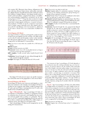
occurs in the AV node, and all impulses conduct to the ventricles, Examples: (A) Second-degree AV block, type I (Wenckebach)
but with delayed conduction times. First-degree AV block can be with 3:2 conduction. (B) Second-degree AV block, type I.
recognized by the following ECG characteristics: Note that the PR interval preceding the pause is longer than
the PR interval after the pause.
Rate: Can occur at any sinus rate, usually 60 to 100 beats per
minute
Rhythm: Regular
V 1
P waves: Normal, precede every QRS
PR interval: Greater than 0.20 second. PR intervals as long as
1 second or more have been reported 22,57 A
QRS complex: Usually normal unless bundle-branch block
exists V 1
Conduction: Normal through the atria, delayed through the AV
node, normal through the ventricles
Example: First-degree AV block (PR interval, 0.44 second) B
The treatment of type I second-degree AV block depends on
the conduction ratio, the resulting ventricular rate, and, most im-
portant, the patient’s tolerance for the rhythm. If the ventricular
rate is slow enough to decrease cardiac output, the treatment is at-
ropine to increase the sinus rate and speed conduction through
the AV node. At higher conduction ratios, where the ventricular
rate is within a normal range, no treatment is necessary. If the
block is due to drug therapy, the drug dose may need to be de-
creased or a pacemaker implanted to control the drug-induced
First-degree AV block does not require any specific treatment,
bradycardia while drug therapy continues. This type of block is
but it should be observed for progression to more serious block.
usually temporary and benign and seldom requires pacing, al-
though temporary pacing may be needed when the ventricular
Second-Degree AV Block
rate is slow.
Second-degree AV block occurs when one atrial impulse at a time
fails to be conducted to the ventricles. Second-degree AV block is Type II. Type II second-degree AV block, also called Mobitz
divided into two categories: type I block, usually occurring in the II, is sudden failure of conduction of an atrial impulse to the ven-
I
I
AV node, and type II block, occurring below the AV node in the tricles without progressive increases in conduction time of consec-
bundle of His or bundle-branch system. utive P waves. Type II block occurs below the AV node and is usu-
ally associated with bundle-branch block; therefore, the dropped
Type I (Wenckebach). Type I second-degree AV block, of- beats are usually a manifestation of bilateral bundle-branch block.
I
I
ten referred to as Wenckebach or Mobitz I, is a progressive increase In this form of block, there is no progressive increase in PR inter-
in conduction times of consecutive atrial impulses into the ventri- vals before the blocked P waves. Type II block is less common but
cles until one impulse fails to conduct, or is “dropped.” This ap- more serious than type I block. Type II second-degree AV block
pears on the ECG as gradually lengthening PR intervals until one can be recognized by the following ECG characteristics:
P wave fails to conduct and is not followed by a QRS complex, re-
sulting in a pause, after which the cycle repeats itself. Rate: Can occur at any basic rate
Type I second-degree AV block can be recognized by the fol- Rhythm: Irregular due to blocked beats unless 2:1 conduction is
lowing ECG characteristics: present