
Page 391 - Cardiac Nursing
P. 391
1
1
2:1
/09
/09
1
M
M
Pa
2:1
6 A
6 A
q
q
xd
87.
87.
q
/30
/30
/09
xd
6
6
Pa
t
ara
ara
p
p
t
In
c.
c.
a
a
In
g
e 3
e 3
Pa
g
g
A
A
p
67
67
67
3-3
p
LWB K34 0-c 16_ pp333-387.qxd 6/30/09 12:16 AM Page 367 Aptara Inc.
33
p
LWBK340-c16_
0-c
16_
LWB
K34
33
3-3
C HAPTER 1 6 / Arrhythmias and Conduction Disturbances 367
Rate: Ventricular rates up to 300 beats per minute waves can simulate left bundle-branch block (LBBB), anterior
Rhythm: Irregular. Often appears as groups of very short R-R in- MI, anterior fascicular block, and left ventricular hypertro-
tervals alternating with groups of longer R-R intervals. The phy. 22,62,65,66
longest R-R intervals are often more than twice the shortest R-
R intervals. Variants of Preexcitation Syndromes
P waves: None, because atria are fibrillating In addition to the Kent bundle described above, which is respon-
PR interval: None sible for WPW syndrome, other anatomical connections exist that
QRS complex: Wide, bizarre due to abnormal depolarization of can bypass the normal AV node delay or create connections be-
ventricles through accessory pathway tween different parts of the conduction system and the ventricles
Conduction: Disorganized and chaotic through atria. Atrial im- and cause variations of the preexcitation pattern. Fibers originat-
pulses conduct into ventricles through accessory pathway, re- ing in the atria and inserting into the His bundle (atriohisian
sulting in muscle cell-to-cell conduction through ventricles. fibers) have been demonstrated anatomically and can result in a
short PR interval and normal QRS complex. This pattern used to
Immediate treatment of AF with anterograde conduction be called Lown–Ganong–Levine syndrome (Fig. 16-10), but evi-
through an accessory pathway depends on ventricular rate and the dence does not support a specific syndrome consisting of short
patient’s tolerance of the arrhythmia. Cardioversion is the treat- PR, normal QRS, and tachycardias that can be proven to be re-
ment of choice when severe hemodynamic impairment occurs. lated to these fibers. 22
Drug treatment is directed at slowing conduction through the ac- Another variant of preexcitation involves conduction over a
cessory pathway and restoring and maintaining sinus rhythm. pathway that originates in either the atrium or the AV node and
Drugs that increase the refractory period and depress conduction inserts into the right bundle branch (atriofascicular or nodofasci-
in the bypass tract include procainamide, flecainide, propafenone, cular fibers, also called Mahaim fibers), resulting in a wide QRS
amiodarone, and sotalol. Many of these drugs are also effective in (usually LBBB morphology). In these variants, the PR interval
preventing recurrences of AF. Digoxin and calcium channel block- may be normal or short. Reentrant tachycardias can occur with
ers, commonly used to treat AF that conducts through the AV any of these variations in anatomy, and the QRS may be normal
node, are contraindicated whenever the tachycardia is due to an- or wide during tachycardia, depending on the location of the ac-
terograde conduction through an accessory pathway because they cessory pathways responsible.
can accelerate conduction through the bypass tract or depress ven-
tricular contractility, leading to hemodynamic deteriora- Treatment
tion. 22,61,64 Preexcitation does not require treatment unless it is associated with
WPW syndrome can resemble other conditions usually diag- symptomatic tachyarrhythmias. Ideally, specific therapy should be
nosed by ECG. The presence of anteriorly directed delta waves based on a known mechanism of the arrhythmia and knowledge of
can simulate RBBB, posterior or inferior MI, right ventricular hy- a drug’s effect on that mechanism in both conduction pathways.
pertrophy, or posterior fascicular block. Posteriorly directed delta This knowledge is best gained through electrophysiologic study,
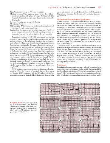
■ Figure 16-10 ECG showing a short
PR interval and normal QRS (formerly
called Lown–Ganong–Levine syndrome).
Upright P waves in inferior leads and neg-
ative P wave in aVR indicate a sinus ori-
gin, not junctional rhythm.