
Page 702 - Cardiac Nursing
P. 702
09
qxd
49
P
AM
5-7
p65
7
04.
/1/
g
K34
0-c
LWB
L L LWB K34 0-c 28_ p65 5-7 04. qxd 7 /1/ 09 9: 49 AM P a a g e e 678 Ap tar a a
LWBK340-c28_p655-704.qxd 7/1/09 9:9:49 AM Page 678 Aptara
Ap
678
tar
28_
678 PA R T I V / Pathophysiology and Management Disease
A
B
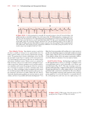
■ Figure 28-24 (A) Safety pacing due to crosstalk: AV sequential pacing at a short AV interval of about 100
milliseconds due to ventricular sensing of the atrial output pulse. (B) Safety pacing due to sensing early in the
AV delay. The first four beats are AV sequential pacing at the programmed AV delay of about 160 milliseconds.
The star beat is the first beat in a run of ventricular tachycardia (VT) that occurs immediately after the atrial
pacing spike. When the ventricular channel “opened its eyes” after the blanking period, it saw the QRS very
early in the AV delay, and rather than inhibit its output, it paced at the safety pacing AV delay of approximately
100 milliseconds. Safety pacing prevents inappropriate inhibition of ventricular pacing but delivers the ven-
tricular output early enough to avoid the T wave.
Rate-Adaptive Pacing. Rate-adaptive pacing is used when likely that future pacemakers will combine two or more sensors to
the heart is unable to increase its rate appropriately when the get the most physiologic response to the body’s needs for increased
body’s need for cardiac output increases (chronotropic incompe- cardiac output. Figures 28-25 and 28-26 illustrate ECG examples
tence). The pacing system contains a physiologic sensor that tells of rate adaptive pacing, which can appear as pacemaker malfunc-
the pacemaker to pace faster in response to the sensed parameter. tion if the observer is unaware of the rate response feature.
The most frequently used sensors at this time are motion sensors
and minute ventilation sensors. Motion sensors are activated by Atrial Overdrive Pacing. Atrial pacing at rapid rates of 200
body movement, such as occurs with exercise, and signal the pace- to 500 impulses/min is used in an attempt to terminate atrial
maker to pace faster. Minute ventilation sensors measure transtho- tachyarrhythmias such as atrial tachycardia, atrial flutter, and
racic impedance and increase the pacing rate when the respiratory atrial fibrillation (Fig. 28-27). This type of pacing is most fre-
rate is increased in response to exercise, emotional states, fever, quently performed using a temporary pulse generator and pacing
and so on. Other technologies being investigated include sensors through epicardial leads in cardiac surgery patients. It can also be
for metabolic parameters like blood temperature and venous oxy- performed with a transvenous atrial lead, but this is less effective.
gen saturation, and sensors of cardiac indices like QT interval, Newer dual-chamber temporary pulse generators have overdrive
ventricular depolarization gradient, pre-ejection interval, stroke pacing capability. It is extremely important to accurately identify
volume, and rate of myocardial wall tension development. 28 It is the atrial pacing wires and make sure that rapid pacing is not
70
Rate 70
Rate 70
Rate 7 70
Rate
Rate
■ Figure 28-25 VVIR pacing. Note the increase in VVI
pacing rate from 70 bpm to 87 bpm with activity.
R R R R R R Rate 75
at at a a a
Ra e e e e te te 7 7 7 7 7 7 5 5 5
Raate 75
ate 87
ate
ate
Ra
R
Ra
Ra e 8 8 8 7 7 7 7 7 7 7
a
a
ate