
Page 703 - Cardiac Nursing
P. 703
7
/1/
09
qxd
p65
5-7
04.
679
Ap
tar
g
49
AM
P
LWB
LWBK340-c28_p655-704.qxd 7/1/09 9:9:49 AM Page 679 Aptara
LWB K34 0-c 28_ p65 5-7 04. qxd 7 /1/ 09 9: 49 AM P a a g e e 679 Ap tar a a
28_
0-c
K34
C HAPTER 2 8 / Pacemakers and Implantable Defibrillators 679
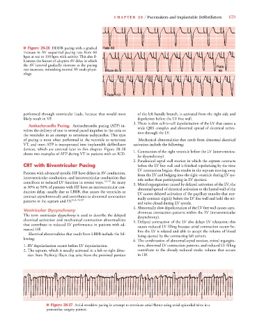
e 60
■ Figure 28-26 DDDR pacing with a gradual Rate 60
ate 6
Rate 60
Rate 60
a
increase in AV sequential pacing rate from 60
bpm at rest to 110 bpm with activity. This also il-
lustrates the feature of adaptive AV delay in which
the AV interval gradually shortens as the pacing Rate
rate increases, mimicking normal AV node physi- Ra a e e e e e e e t t t at at ate 8 85 96
Ra
Ra
5 5 5
5
85
R R R R R R R R R Rate 85
8 8 8 8 8 8
85
ology.
R Ra
R R R R R R Rate 1 1 1 1 11 1 1 1 110
Ra
0 0 0 0 0 0 00
11
e 1
11
e e e e e t t at at a a
performed through ventricular leads, because that would most of the left bundle branch, is activated from the right side and
likely result in VF. depolarizes before the LV free wall.
3. There is slow cell-to-cell depolarization of the LV that causes a
Antitachycardia Pacing. Antitachycardia pacing (ATP) in- wide QRS complex and abnormal spread of electrical activa-
volves the delivery of one to several paced impulses to the atria or tion through the LV.
the ventricles in an attempt to terminate tachycardias. This type
of pacing is most often performed in the ventricle to terminate Mechanical abnormalities that result from abnormal electrical
VT, and most ATP is incorporated into implantable defibrillator activation include the following:
devices, which are covered later in this chapter. Figure 28-28 1. Contraction of the right ventricle before the LV (interventricu-
shows two examples of ATP during VT in patients with an ICD.
lar dysynchrony).
2. Paradoxical septal wall motion in which the septum contracts
CRT with Biventricular Pacing before the LV free wall and is finished repolarizing by the time
LV contraction begins; this results in the septum moving away
Patients with advanced systolic HF have delays in AV conduction, from the LV and bulging into the right ventricle during LV sys-
interventricular conduction, and intraventricular conduction that tole rather than participating in LV ejection.
contribute to reduced LV function in several ways. 1,6–9 As many 3. Mitral regurgitation caused by delayed activation of the LV; the
as 30% to 50% of patients with HF have an intraventricular con- abnormal spread of electrical activation to the lateral wall of the
duction delay, usually due to LBBB, that causes the ventricles to LV causes delayed activation of the papillary muscles that nor-
contract asynchronously and contributes to abnormal contraction mally contract slightly before the LV free wall and hold the mi-
patterns in the septum and LV. 8,9,11,29
tral valve closed during LV systole.
4.Abnormally slow depolarization of the LV free wall causes asyn-
Ventricular Dysynchrony chronous contraction patterns within the LV (intraventricular
The term ventricular dysynchrony is used to describe the delayed dysynchrony).
electrical activation and mechanical contraction abnormalities 5. Delayed contraction of the LV also delays LV relaxation; this
that contribute to reduced LV performance in patients with ad- causes reduced LV filling because atrial contraction occurs be-
vanced HF. fore the LV is relaxed and able to accept the volume of blood
Electrical abnormalities that result from LBBB include the fol- being ejected by the contracting left atrium.
lowing:
6. The combination of abnormal septal motion, mitral regurgita-
1. RV depolarization occurs before LV depolarization. tion, abnormal LV contraction patterns, and reduced LV filling
2. The septum, which is usually activated in a left-to-right direc- contribute to the already reduced stroke volume that occurs
tion from Purkinje fibers that arise from the proximal portion in HF.
■ Figure 28-27 Atrial overdrive pacing in attempt to terminate atrial flutter using atrial epicardial wires in a
postcardiac surgery patient.