
Page 285 - ACCCN's Critical Care Nursing
P. 285
262 P R I N C I P L E S A N D P R A C T I C E O F C R I T I C A L C A R E
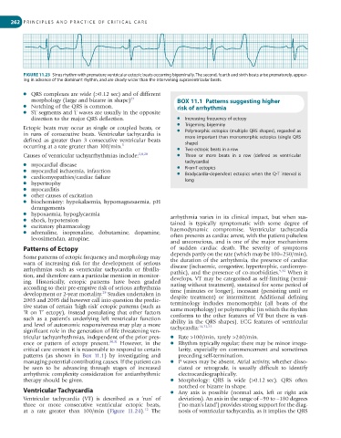
FIGURE 11.23 Sinus rhythm with premature ventricular ectopic beats occurring bigeminally. The second, fourth and sixth beats arise prematurely, appear-
ing in advance of the dominant rhythm, and are clearly wider than the intervening supraventricular beats.
● QRS complexes are wide (>0.12 sec) and of different
morphology (large and bizarre in shape) 27 BOX 11.1 Patterns suggesting higher
● Notching of the QRS is common. risk of arrhythmia
● ST segments and T waves are usually in the opposite
direction to the major QRS deflection. ● Increasing frequency of ectopy
● Trigeminy, bigeminy
Ectopic beats may occur as single or coupled beats, or ● Polymorphic ectopics (multiple QRS shapes), regarded as
in runs of consecutive beats. Ventricular tachycardia is more important than monomorphic ectopics (single QRS
defined as greater than 3 consecutive ventricular beats shape)
occurring at a rate greater than 100/min. 5 ● Two ectopic beats in a row
Causes of ventricular tachyarrhythmias include: 3,8,28 ● Three or more beats in a row (defined as ventricular
tachycardia)
● myocardial disease ● R-on-T ectopics
● myocardial ischaemia, infarction ● Bradycardia-dependent ectopics when the Q-T interval is
● cardiomyopathies/cardiac failure long
● hypertrophy
● myocarditis
● other causes of excitation
● biochemistry: hypokalaemia, hypomagnesaemia, pH
derangements
● hypoxaemia, hypoglycaemia arrhythmia varies in its clinical impact, but when sus-
● shock, hypotension tained is typically symptomatic with some degree of
● excitatory pharmacology haemo dynamic compromise. Ventricular tachycardia
● adrenaline, isoprenaline, dobutamine, dopamine, often presents as cardiac arrest, with the patient pulseless
levosimendan, atropine.
and unconscious, and is one of the major mechanisms
Patterns of Ectopy of sudden cardiac death. The severity of symptoms
Some patterns of ectopic frequency and morphology may depends partly on the rate (which may be 100–250/min),
the duration of the arrhythmia, the presence of cardiac
warn of increasing risk for the development of serious disease (ischaemic, congestive, hypertrophic, cardiomyo-
arrhythmias such as ventricular tachycardia or fibrilla- pathic), and the presence of co-morbidities. 9,32 When it
tion, and therefore earn a particular mention in monitor- develops, VT may be categorised as self-limiting (termi-
ing. Historically, ectopic patterns have been graded nating without treatment), sustained for some period of
according to their pre-emptive risk of serious arrhythmia time (minutes or longer), incessant (persisting until or
29
development or 2-year mortality. Studies undertaken in despite treatment) or intermittent. Additional defining
2003 and 2005 did however call into question the predic- terminology includes monomorphic (all beats of the
tive status of certain ‘high risk’ ectopic patterns (such as same morphology) or polymorphic (in which the rhythm
‘R on T’ ectopy), instead postulating that other factors conforms to the other features of VT but there is vari-
such as a patient’s underlying left ventricular function ability in the QRS shapes). ECG features of ventricular
and level of autonomic responsiveness may play a more tachycardia: 14,32,33
significant role in the generation of life threatening ven-
tricular tachyarrhythmias, independent of the prior pres- ● Rate >100/min, rarely >240/min.
ence or pattern of ectopy present. 30,31 However, in the ● Rhythm typically regular; there may be minor irregu-
critical care context it is reasonable to respond to certain larity, especially on commencement and sometimes
patterns (as shown in Box 11.1) by investigating and preceding self-termination.
managing potential contributing causes. If the patient can ● P waves may be absent. Atrial activity, whether disso-
be seen to be advancing through stages of increased ciated or retrograde, is usually difficult to identify
arrhythmic complexity consideration for antiarrhythmic electrocardiographically.
therapy should be given. ● Morphology: QRS is wide (>0.12 sec). QRS often
notched or bizarre in shape.
Ventricular Tachycardia ● Any axis is possible (normal axis, left or right axis
Ventricular tachycardia (VT) is described as a ‘run’ of deviation). An axis in the range of −90 to −180 degrees
three or more consecutive ventricular ectopic beats, (‘no man’s land’) provides strong support for the diag-
12
at a rate greater than 100/min (Figure 11.24). The nosis of ventricular tachycardia, as it implies the QRS