
Page 284 - ACCCN's Critical Care Nursing
P. 284
Cardiac Rhythm Assessment and Management 261
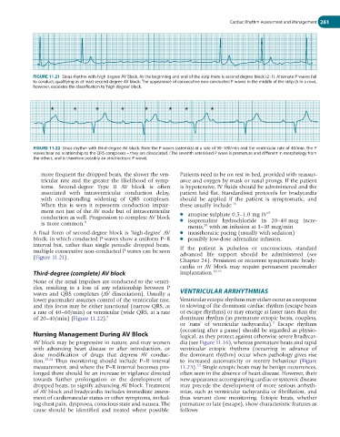
FIGURE 11.21 Sinus rhythm with high degree AV Block. At the beginning and end of the strip there is second degree block (2 : 1). Alternate P waves fail
to conduct, qualifying as at least second degree AV block. The appearance of consecutive non-conducted P waves in the middle of the strip (5 in a row),
however, escalates the classification to ‘high degree’ block.
* * * * * * * *
FIGURE 11.22 Sinus rhythm with third-degree AV block. Note the P waves (asterisks) at a rate of 90–100/min and the ventricular rate of 40/min. The P
waves bear no relationship to the QRS complexes – they are dissociated. (The seventh asterisked P wave is premature and different in morphology from
the others, and is therefore possibly an atrial ectopic P wave).
more frequent the dropped beats, the slower the ven- Patients need to be on rest in bed, provided with reassur-
tricular rate and the greater the likelihood of symp- ance and oxygen by mask or nasal prongs. If the patient
toms. Second-degree Type II AV block is often is hypotensive, IV fluids should be administered and the
asso ciated with intraventricular conduction delay, patient laid flat. Standardised protocols for bradycardia
with corresponding widening of QRS complexes. should be applied if the patient is symptomatic, and
When this is seen it represents conduction impair- these usually include: 18
ment not just of the AV node but of intraventricular ● atropine sulphate 0.5–1.0 mg IV 25
conduction as well. Progression to complete AV block ● isoprenaline hydrochloride in 20–40 mcg incre-
is more common. 9
26
ments, with an infusion at 1–10 mcg/min
A final form of second-degree block is ‘high-degree’ AV ● transthoracic pacing (usually with sedation)
block, in which conducted P waves show a uniform P–R ● possibly low-dose adrenaline infusion.
interval but, rather than single periodic dropped beats,
multiple consecutive non-conducted P waves can be seen If the patient is pulseless or unconscious, standard
(Figure 11.21). advanced life support should be administered (see
Chapter 24). Persistent or recurrent symptomatic brady-
cardia or AV block may require permanent pacemaker
Third-degree (complete) AV block implantation. 18,19
None of the atrial impulses are conducted to the ventri-
cles, resulting in a loss of any relationship between P
waves and QRS complexes (AV dissociation). Usually a VENTRICULAR ARRHYTHMIAS
lower pacemaker assumes control of the ventricular rate, Ventricular ectopic rhythms may either occur as a response
and this focus may be either junctional (narrow QRS, at to slowing of the dominant cardiac rhythm (escape beats
a rate of 40–60/min) or ventricular (wide QRS, at a rate or escape rhythms) or may emerge at faster rates than the
of 20–40/min) (Figure 11.22). 9 dominant rhythm (as premature ectopic beats, couplets,
9
or ‘runs’ of ventricular tachycardia). Escape rhythms
(occurring after a pause) should be regarded as physio-
Nursing Management During AV Block logical, as they protect against otherwise severe bradycar-
AV block may be progressive in nature, and may worsen dia (see Figure 11.16), whereas premature beats and rapid
with advancing heart disease or after introduction, or ventricular ectopic rhythms (occurring in advance of
dose modification of drugs that depress AV conduc- the dominant rhythm) occur when pathology gives rise
tion. 23,24 Thus monitoring should include P–R interval to increased automaticity or reentry behaviour (Figure
7,9
measurement, and where the P–R interval becomes pro- 11.23). Single ectopic beats may be benign occurrences,
longed there should be an increase in vigilance directed often seen in the absence of heart disease. However, their
towards further prolongation or the development of new appearance accompanying cardiac or systemic disease
dropped beats, to signify advancing AV block. Treatment may precede the development of more serious arrhyth-
of AV block and bradycardia includes immediate assess- mias, such as ventricular tachycardia or fibrillation, and
ment of cardiovascular status or other symptoms, includ- thus warrant close monitoring. Ectopic beats, whether
ing chest pain, dyspnoea, conscious state and nausea. The premature or late (escape), show characteristic features as
cause should be identified and treated where possible. follows: