
Page 1569 - Hematology_ Basic Principles and Practice ( PDFDrive )
P. 1569
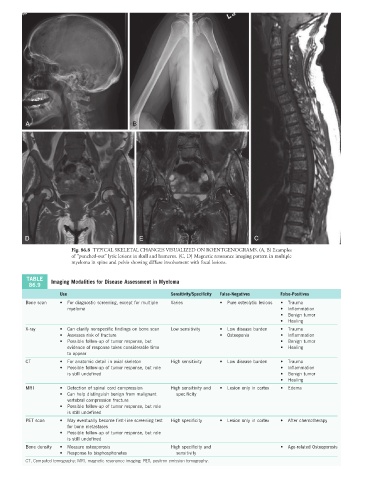
A                                  B


         D                                     E                                    C
                        Fig. 86.8  TYPICAL SKELETAL CHANGES VISUALIZED ON ROENTGENOGRAMS. (A, B) Examples
                        of “punched-out” lytic lesions in skull and humerus. (C, D) Magnetic resonance imaging pattern in multiple
                        myeloma in spine and pelvis showing diffuse involvement with focal lesions.

          TABLE   Imaging Modalities for Disease Assessment in Myeloma
          86.9
                     Use                                 Sensitivity/Specificity  False-Negatives  False-Positives
         Bone scan   •  For diagnostic screening, except for multiple   Varies  •  Pure osteolytic lesions  •  Trauma
                       myeloma                                                               •  Inflammation
                                                                                             •  Benign tumor
                                                                                             •  Healing
         X-ray       •  Can clarify nonspecific findings on bone scan  Low sensitivity  •  Low disease burden  •  Trauma
                     •  Assesses risk of fracture                        •  Osteopenia       •  Inflammation
                     •  Possible follow-up of tumor response, but                            •  Benign tumor
                       evidence of response takes considerable time                          •  Healing
                       to appear
         CT          •  For anatomic detail in axial skeleton  High sensitivity  •  Low disease burden  •  Trauma
                     •  Possible follow-up of tumor response, but role                       •  Inflammation
                       is still undefined                                                    •  Benign tumor
                                                                                             •  Healing
         MRI         •  Detection of spinal cord compression  High sensitivity and   •  Lesion only in cortex  •  Edema
                     •  Can help distinguish benign from malignant   specificity
                       vertebral compression fracture
                     •  Possible follow-up of tumor response, but role
                       is still undefined
         PET scan    •  May eventually become first-line screening test   High specificity  •  Lesion only in cortex  •  After chemotherapy
                       for bone metastases
                     •  Possible follow-up of tumor response, but role
                       is still undefined
         Bone density  •  Measure osteoporosis           High specificity and                •  Age-related Osteoporosis
                     •  Response to bisphosphonates        sensitivity
         CT, Computed tomography; MRI, magnetic resonance imaging; PET, positron emission tomography.