
Page 899 - Hematology_ Basic Principles and Practice ( PDFDrive )
P. 899
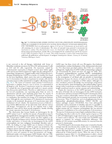
782    Part VII  Hematologic Malignancies


                                                  Reactivation of  Random X              Polyclonal
                                                    paternal X   inactivation            proliferation


                          Oocyte


                         Sperm                Female       Morula          Blastocyst
                                               zygote

                                                                                         Monoclonal
                                                                                         proliferation
                        Fig.  56.7  X-CHROMOSOME–LINKED ENZYME GLUCOSE-6-PHOSPHATE DEHYDROGENASE
                        (G6PD) AS A MARKER TO INVESTIGATE CLONAL DEVELOPMENT OF HUMAN HEMATOPOI-
                        ETIC DISORDERS. Early in embryogenesis, regions of all but one X chromosome are inactivated in each
                        cell  containing  two  or  more  X  chromosomes. The  choice  of  maternal  versus  paternal  X  chromosome  for
                        inactivation is random. Once the inactivation occurs, it is fixed and is stably transmitted to daughter cells
                        during mitosis (Lyon hypothesis). Females who are heterozygous for the common B type and the less frequent
                        A type, G6PD (localized on Xq27), are mosaic. This cellular mosaicism is used to study monoclonal versus
                        polyclonal cell proliferation and development of malignant hematopoietic diseases. (Courtesy Dr. W Raskind,
                        University of Washington, Seattle.)


        is  not  restricted  to  the  cell  lineages.  Individuals  with  Turner  or   G6PD type, but these clonal cells were Ph-negative; thus leukemic
        Klinefelter syndrome are mosaic for XX or XY and monosomy X cells   transformation predates development of the chromosomal abnormal-
        or  XXY  and  XY  cells,  respectively.  The  mosaicism  created  by   ity. This  observation  provided  evidence  that  CML  has  a  multistep
        X-chromosome inactivation in females is much more widely appli-  pathogenesis. Application of G6PD studies to hematologic malignan-
        cable and has provided fundamental insights into the pathogenesis of   cies  demonstrated  the  clonal  and  stem  cell  origin  for  AML,  ALL,
        hematologic malignancies. Original studies with X-linked glucose-6-  Ph-negative  myeloproliferative  neoplasm  (MPN),  myelodysplastic
        phospate dehydrogenase (G6PD) as a marker of clonality were based   syndrome (MDS), and CLL. G6PD studies were particularly useful
        on the Lyon hypothesis, which asserts that early in embryogenesis,   in the investigation of red blood cells and platelets in hematologic
        one X chromosome in females is inactivated in somatic cells and the   malignancies because the absence of nuclei in these cells means they
        activation status is stably transmitted to daughter cells during mitosis   cannot be studied by cytogenetics or DNA analysis. Although it is
        (Fig. 56.7). The choice of maternal versus paternal X-chromosome   now considered common knowledge that hematologic malignancies
        inactivation is random; however, once it occurs, it is maintained in   are characterized by clonal development, this understanding is greatly
        all daughter cells. Random X inactivation occurs by embryonic day   owing to what is now known as classic Fialkow’s work, whose profound
        6.5 around the start of gastrulation and results in a mosaic pattern   insight contributed much to current concepts and understanding.
        that characterizes adult females. Therefore an adult female is a mosaic   Despite the importance of the G6PD approach, it is limited by
        for  two-cell  populations,  one  expressing  genes  from  an  active  X   the rarity of females who are heterozygous for the G6PD isoenzyme.
        chromosome  and  the  other  expressing  genes  from  the  inactive  X   An alternative and more extensive DNA-based X-chromosome clonal
        chromosome. Incidentally, mammalian X-chromosome inactivation   assay uses common polymorphic markers that are caused by changes
        is a mechanism that equalizes the dosage of X-linked genes between   in DNA methylation patterns that accompany inactivation of the X
        sexes. Although the exact mechanism of X-chromosome inactivation   chromosome. These X-linked loci, such as phosphoglycerine kinase,
        remains to be elucidated, the process of X inactivation starts with   hypoxanthine  phosphoribosyltransferase,  DXS25  (M27β),  and
        methylation of CpG islands. The inactivation process is believed to   human  androgen  receptor  (HUMARA),  have  been  subsequently
        occur before differentiation of the embryonic stem cell into various   extensively used in assessment of clonality, and now it is possible to
        cell  lineages.  Hematopoietic  cells  do  not  originate  from  a  single   identify clonal cell populations in virtually all females. DNA-based
        embryonic stem cell but from several progenitors, thereby allowing   marker systems rely on a sequence polymorphism that has adjacent
        for mosaic expression from both X chromosomes.        differences in methylation on the active and inactive X chromosomes.
           The  observation  that  human  females  are  heterozygous  for  the   The inactive X chromosome is more highly methylated than its active
                         −
        G6PD variant A and A  and that two mosaic cell populations may be   homologue, but this is only true for certain regions of genes as 10%
        distinguishable by electrophoretic mobility was reported in the 1960s.   to 20% of X-linked genes escape inactivation and can be found both
        The  X-inactivation  G6PD  mosaic  system  was  then  applied  to  the   in clusters and in isolation. The most widely used HUMARA assay
        study of clonality in human tumors (uterine leiomyomas) in 1964 by   appears  to  maintain  the  stringent  methylation  differences.  The
        Gartler and Linden. In females who are heterozygous for the G6PD   number of CAG tandem repeats differentiates the maternal from the
        polymorphism  and  have  malignant  hematologic  disorders  such  as   paternal X chromosome.
        CML, the finding of a single G6PD type in marrow or blood cells   The DNA-based X-chromosome clonal assay is limited to females
        and  both  the  A  and  B  type  G6PD  in  tissues  not  involved  by  the   younger than 60 years because they usually have 1 : 1 distribution of
        malignant process demonstrated that CML was of clonal origin and   two-mosaic–cell  population.  A  ratio  greater  than  3 : 1  is  found  in
        provided evidence that the malignant transformation occurred at the   women older than 60 years, probably as a result of stem cell kinetics
        level  of  a  stem  cell  common  to  most  hematopoietic  cell  lineages.   influenced by X-linked genetic factors. When the ratio of two cell
        Additional studies with heterozygous G6PD females who had CML   populations is greater than 3 : 1, this phenomenon is called a skewed
        demonstrated  that some CML-derived  B  lymphocytes  had  a single   X-inactivation pattern. With the HUMARA assay, acquired unequal